Hypodermis (Subcutaneous Tissue) Lesson: Function & Structure
Lesson Overview
The hypodermis plays a crucial role in regulating body temperature by insulating the body and storing fat for energy. Also, it serves as a protective cushion for underlying tissues and organs. Understanding the formation of the hypodermis is essential to grasping the health and function of your skin.
What Is the Hypodermis?
The hypodermis, also known as subcutaneous tissue, is the deepest of the three main layers of your skin. These layers are the epidermis (outermost), dermis (middle), and hypodermis (innermost). The hypodermis lies directly beneath the dermis and connects the skin to underlying tissues like muscles and bone. Composed primarily of loose connective and adipose (fat) tissue, its thickness and composition vary across the body.
For example, the hypodermis is typically thicker in the thighs and abdomen compared to the scalp.
Where Is the Hypodermis Located?
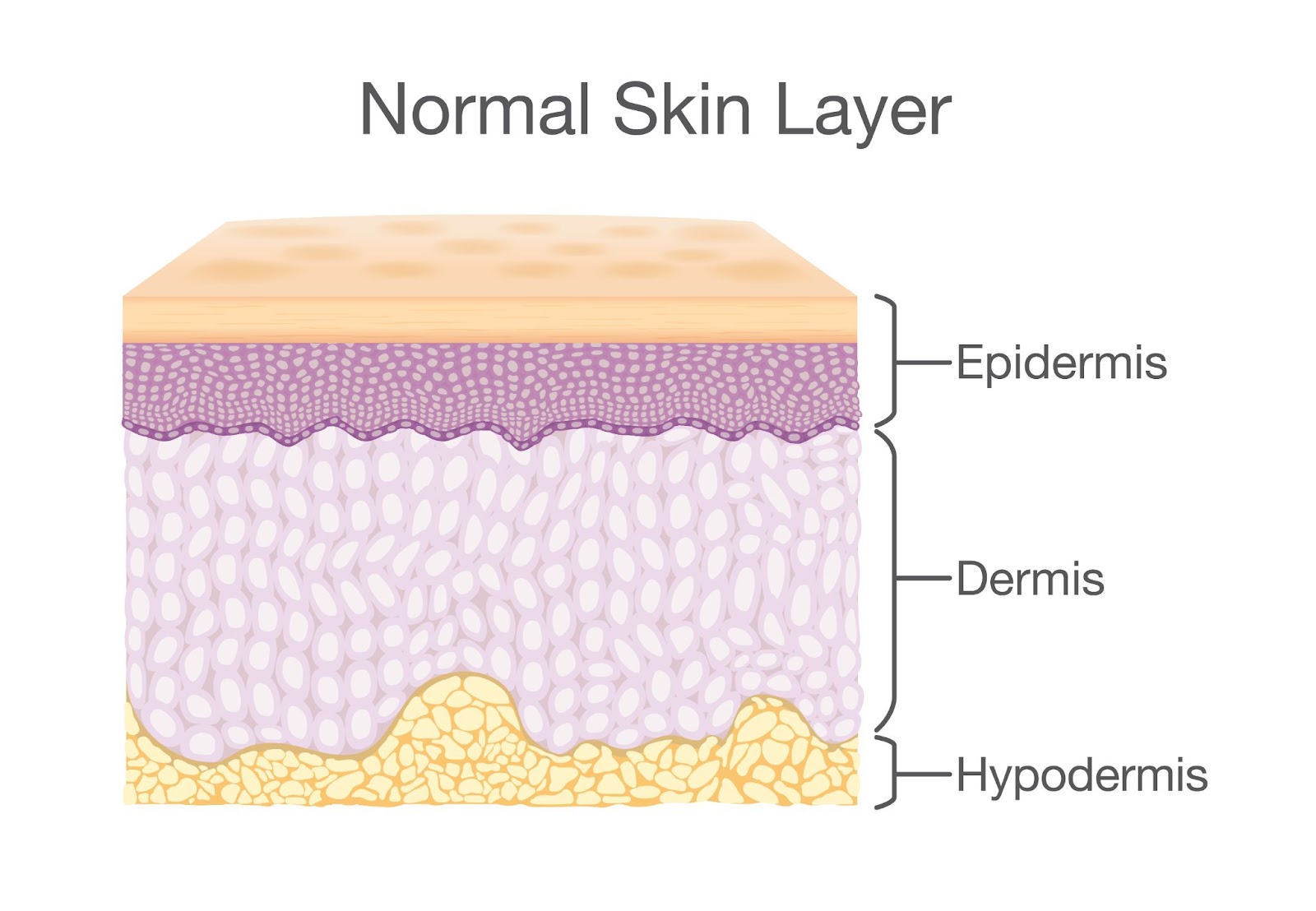
Fig. shows the three layers of skin: the epidermis is the outermost layer, the dermis is the thicker middle layer with a wavy boundary, and the hypodermis is the bottom layer with a distinct yellow fatty layer.
- Location: Deepest layer of the skin, beneath the dermis.
- Not Part of the Skin: Not technically part of the skin itself, but a layer of tissue beneath it.
- Connection: Serves as a bridge between the skin and underlying structures.
- Position Relative to Fascia: Located superficial (above) to the fascia (fibrous connective tissue surrounding muscles, bones, etc.). Positioned between the dermis and the fascia.
- Boundary with Fascia: Connection to fascia is a gradual transition, not a sharp boundary.
- Thickness Variation: Thickness varies across body regions and between individuals. Thicker in areas needing cushioning/insulation (e.g., abdomen, buttocks, thighs) and in individuals with a higher body fat percentage. Thinner in areas like the scalp and back of hands.
Structure and Components of the Hypodermis
The hypodermis is a complex layer with distinct structural components that contribute to its diverse functions. Its primary constituents are loose connective tissue and adipose tissue (fat).
1. Loose Connective Tissue: This tissue forms a network of fibers that provide support and structure to the hypodermis. It's characterized by its loosely arranged fibers, creating spaces that allow for flexibility and the passage of blood vessels, nerves, and lymphatic vessels. Think of it as the framework that holds everything together, but with enough give for movement and growth.
2. Adipose Tissue (Fat): Adipose tissue is the dominant component of the hypodermis, particularly in certain areas of the body. It's composed of specialized cells called adipocytes, which are filled with fat droplets. This fat serves several crucial functions, including insulation, energy storage, and cushioning. The amount and distribution of adipose tissue contribute significantly to body contour and vary between individuals due to factors like genetics, hormones, and diet. For example, the hypodermis in the abdomen and thighs tends to have a higher concentration of adipose tissue compared to the face or scalp.
3. Blood Vessels: The hypodermis contains a network of blood vessels that supply the skin with nutrients and oxygen, and also help regulate body temperature. These vessels branch off from larger vessels in deeper tissues and form a complex network throughout the hypodermis.
4. Nerves: Nerves in the hypodermis transmit sensory information, such as touch, pressure, temperature, and pain. These nerves play a crucial role in our interaction with the environment.
5. Lymphatic Vessels: Lymphatic vessels are also present in the hypodermis, and they play a role in the immune system by collecting fluid, waste products, and immune cells.
6. Other Components: While less abundant, the hypodermis also contains other components like hair follicle roots, sweat glands, and sensory receptors.
The relative proportions of these components, particularly loose connective tissue and adipose tissue, vary depending on the body region and individual. For instance, the hypodermis in the palms of the hands and soles of the feet contains more connective tissue for added strength and protection, while the hypodermis in the abdomen and buttocks has a higher proportion of adipose tissue for insulation and energy storage. This regional variation reflects the specialized functions of the hypodermis in different parts of the body.
Take This Quiz :

Functions of the Hypodermis
The hypodermis performs several crucial functions, contributing significantly to overall skin health and body homeostasis. These functions are primarily attributed to its composition of loose connective tissue and adipose (fat) tissue.
1. Insulation: The abundant adipose tissue in the hypodermis acts as an insulator, helping to minimize heat loss and maintain body temperature. Fat is a poor conductor of heat, effectively slowing down the transfer of heat from the body's core to the external environment. This is particularly important in colder climates or when the body needs to conserve energy. For example, the thicker hypodermis in individuals with a higher body fat percentage provides greater insulation, helping them to stay warm.
2. Energy Storage: Adipose tissue serves as a major energy reserve. Fat stored in adipocytes can be broken down and used as fuel when the body requires additional energy, such as during periods of fasting, prolonged exercise, or illness. This stored energy is crucial for survival and allows the body to function even when food intake is limited. For example, during prolonged starvation, the body utilizes stored fat reserves in the hypodermis to meet its energy needs.
3. Protection: The hypodermis provides a cushioning layer that protects underlying structures, such as muscles, bones, and internal organs, from physical trauma. The adipose tissue absorbs and dissipates mechanical forces, acting like a shock absorber. This is particularly important in areas prone to impact, such as the buttocks and the soles of the feet. For example, a fall onto the buttocks is less likely to cause serious injury due to the protective cushioning provided by the hypodermis.
4. Connection: The hypodermis connects the skin to underlying tissues, including muscles and bones. This connection is facilitated by the network of connective tissue fibers that extend from the hypodermis into the deeper tissues. This anchoring function helps to stabilize the skin and prevent it from moving excessively. For example, the hypodermis helps to keep the skin taut and prevents it from sagging.
5. Body Contouring: The distribution and amount of adipose tissue in the hypodermis contribute significantly to body shape and contour. Differences in fat distribution between individuals, influenced by factors like genetics and hormones, result in variations in body shape. For example, women tend to have a greater accumulation of fat in the hips and thighs, while men tend to accumulate more fat in the abdomen.
6. Thermoregulation: While the insulating function of fat helps to conserve heat, the hypodermis also plays a role in thermoregulation through its blood vessels. These vessels can dilate (widen) to increase blood flow to the skin surface, allowing for heat dissipation, or constrict (narrow) to reduce blood flow and conserve heat. This vascular response is crucial for maintaining a stable body temperature. For example, during exercise, blood flow to the skin increases, allowing for heat loss and preventing overheating.
7. Site for Injections: Due to its accessibility and relatively low nerve density, the hypodermis is a common site for subcutaneous injections of medications. The rich blood supply in the hypodermis allows for relatively rapid absorption of the injected medication. For example, insulin is often administered via subcutaneous injection.
Differences Between the Epidermis, Dermis, and Hypodermis
The skin, our largest organ, is composed of three distinct layers: the epidermis, dermis, and hypodermis. While they work together to perform the skin's many functions, each layer has unique characteristics in terms of structure, composition, and function.
| Feature | Epidermis | Dermis | Hypodermis (Subcutaneous Tissue) |
| Location | Outermost layer | Middle layer | Deepest layer |
| Composition | Stratified squamous epithelium | Connective tissue (collagen, elastin), blood vessels, nerves, glands | Loose connective tissue, adipose (fat) tissue |
| Blood Vessels | Absent | Present (extensive network) | Present (larger vessels) |
| Nerves | Sparse | Present (sensory and motor) | Present |
| Function | Barrier, protection (UV, pathogens, water loss), vitamin D synthesis | Support, strength, elasticity, temperature regulation, sensation, wound healing | Insulation, energy storage, cushioning, connection to underlying tissues |
| Thickness | Thinnest layer (variable, e.g., thicker on palms/soles) | Thickest layer | Variable (thickest in areas like abdomen, buttocks; thinner on scalp, hands) |
| Other Structures | Melanocytes (pigment cells), keratinocytes | Hair follicles, sweat glands, sebaceous glands | Larger blood vessels and nerves, some sensory receptors |
Rate this lesson:
 Back to top