1.
Which of the following eyelid margin signs are indicative of a case of blepharitis?
Correct Answer
C. Keratinization
Explanation
When inspecting the lid margin, eyecare practitioners should look for vascularization, keratinization, as well as the position of the mucocutaneous junction and punctum. These indications may point to a possible case of blepharitis.1
Reference 1. Jackson WB. Blepharitis: current strategies for diagnosis and management. Can J Ophthalmol. 2008;43:170-9.
2.
All patients with blepharitis are symptomatic.
Correct Answer
B. False
Explanation
Many times, although blepharitis is detectable through clinical signs, patients are asymptomatic. Patients with meibomian gland dysfunction, a common form of posterior blepharitis, frequently have a chronic, low grade lid disease process, often persisting for months or years without producing symptoms (or producing only transitory symptoms when subject to environmental stress). Then, some event or activity may exacerbate the condition and produce more frequent and troubling symptoms.1Reference 1. Jackson WB. Blepharitis: current strategies for diagnosis and management. Can J Ophthalmol. 2008;43:170-9.
3.
Posterior blepharitis should be considered in the differential diagnosis of patients with the symptoms of dry eye.
Correct Answer
A. True
Explanation
Lemp and colleagues studied 159 patients with dry eye symptoms and found that 86% had signs of evaporative dry eye caused by posterior blepharitis/meibomian gland dysfunction.2 Reference 2. Lemp MA, Crews LA, Brown AJ, Foulks GN, Sullivan BD. Distribution of aqueous-deficient and evaporative dry eye in a clinic-based patient cohort: a retrospective study. Cornea. 2012;31:472-8.
4.
Symptoms of blepharitis can be affected by:
Correct Answer
D. All of the above
Explanation
Symptoms not only vary in severity from patient to patient, but can also be affected by seasonal, dietary, or lifestyle changes that are often difficult for a patient to pinpoint. In general, eyecare practitioners must be cognizant of the waxing and waning nature of these conditions, and remember that symptoms may change from season to season, day to day – and even hour to hour.1
Reference
1. Jackson WB. Blepharitis: current strategies for diagnosis and management. Can J Ophthalmol. 2008;43:170-9.
5.
The appearance of normal, healthy meibomian glands is marked by:
Correct Answer
C. Both A and B
Explanation
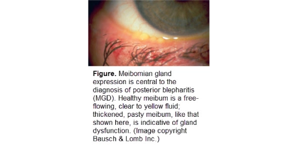
When examining a patient’s eyelid, many eyecare practitioners work anterior to posterior. The first step is to evert the lids, checking for concretions, other lesions, or signs of chronic inflammation – which may or may not be related to blepharitis. Close attention should also be paid to the quality of the lid margin.1 Then, an inspection of the meibomian glands will determine whether the orifices are visible. The appearance of normal, healthy meibomian glands is marked by their location at regular intervals and by a clearly defined cuff of epithelial tissue around each orifice. It is important to check whether the glands appear small, irregular or atrophied, or, conversely, whether they are visibly plugged or capped. Most importantly, the glands must be expressed to grade meibomian gland function (Figure).3
References 1. Jackson WB. Blepharitis: current strategies for diagnosis and management. Can J Ophthalmol. 2008;43:170-9. 4. Viso E, Rodriguez-Ares MT, Abelenda D, Oubina B, Gude F. Prevalence of asymptomatic and symptomatic meibomian gland dysfunction in the general population of Spain. Invest Ophthalmol Vis Sci. 2012;53:2601-6.
6.
Which of the following eyelid signs can indicate blepharitis?
Correct Answer
D. All of the above
Explanation
Upon examination, patients who describe eyelid stickiness and burning eyes upon waking often present with other signs of blepharitis. Patients with anterior blepharitis report a gamut of symptoms. Some patients have red and swollen lids; others complain of irritation and burning. A close look at the lids forms a key part of the examination. Patients with anterior blepharitis often have reddened, swollen lids, telangiectasia, and debris or collarettes along the lashes.1 In posterior blepharitis, the tear meniscus may be foamy, a result of bacterial lipases causing breakdown of the meibomian lipids (Figure).1 In addition, we often see plugged, pouting meibomian glands that yield turbid, viscous meibum – or no meibum at all.1
Reference 1. Jackson WB. Blepharitis: current strategies for diagnosis and management. Can J Ophthalmol. 2008;43:170-9.