Animal Cell Lesson: Structure, Function, and Organelles

Reviewed by Lindsey Block
Lindsey Block, PhD (Cellular & Molecular Biology) |
Lecturer | Molecular Biologist | Reproductive Scientist
Review Board Member
Lindsey, Ph.D. in cellular and molecular biology from the University of Wisconsin-Madison, specializes in Zika's impact on conception and preterm birth biomarkers. She completed courese on Advanced Cell Biology at the University of Illinois at Urbana-Champaign and Advanced Virology at University College Cork. Lindsey's accolades include three first-author papers, three fellowships, and active participation in five conference presentations. Currently associated with the University of Pennsylvania through a T32 NIH Postdoctoral Fellowship, she continues to contribute significantly to her field, combining academic rigor with practical research to advance understanding in reproductive health and prenatal care. Currently, she is a full time lecturer at Northwestern University - The Feinberg School of Medicine.
, PhD (Cellular & Molecular Biology)
Created by ProProfs Editorial Team
The editorial team at ProProfs Quizzes consists of a select group of subject experts, trivia writers, and quiz masters who have authored over 10,000 quizzes taken by more than 100 million users. This team includes our in-house seasoned quiz moderators and subject matter experts. Our editorial experts, spread across the world, are rigorously trained using our comprehensive guidelines to ensure that you receive the highest quality quizzes.
Learn about Our Editorial Process

Lesson Overview

Introduction to Animal Cell Lesson

All animals and plants are composed of many tiny cells. These cells come together to form the structure of every living thing. Whether it's a tree or a dog, cells make up their bodies. Understanding animal cells is essential for studying biology and the various processes that sustain life in animals. This knowledge forms the foundation for exploring more complex biological systems and their interactions within the animal kingdom.

This animal cells lesson will delve into the unique structures and functions of animal cells, focusing on key components such as the cell membrane, nucleus, and mitochondria, which distinguish them from plant cells. Additionally, the lesson will cover the different types of animal cells, their specific roles in the body, and the historical advancements in cell biology that have deepened our understanding of these vital units.

By the end of this lesson, students will gain a comprehensive understanding of animal cell anatomy, the differences between animal and plant cells, and the crucial importance of animal cells in the broader context of life sciences.

What Is an Animal Cell?

An animal cell is the fundamental structural and functional unit of animals, serving as the building block for all animal life. Unlike plant cells, animal cells are characterized by a flexible plasma membrane, which lacks a rigid cell wall, allowing them to adopt various shapes and forms necessary for their diverse functions. This flexibility is crucial for the dynamic activities animal cells perform, such as movement and communication within tissues.

Animal cells contain a variety of specialized organelles, including the nucleus, which houses genetic material and controls cellular activities. Another key organelle is the mitochondrion, known as the powerhouse of the cell, which generates the energy needed to fuel cellular processes. Animal cells also possess smaller vacuoles compared to plant cells, which are involved in storage and transport within the cell.

Typically, animal cells are more varied in shape, reflecting their roles in different tissues, such as muscle contraction, nerve signal transmission, and immune response. The absence of chloroplasts in animal cells distinguishes them from plant cells, as animal cells rely on consuming organic material for energy rather than photosynthesis. The unique composition and organization of animal cells enable them to perform a wide array of vital functions, making them essential to the life and growth of animals.

Fig: Neurons Cells From the Brain Under the Microscope

What Is the History and Development of Cell Theory?

The history and development of cell theory is a fascinating journey that spans several centuries, marked by significant discoveries and contributions from numerous scientists. This timeline provides a detailed account of how cell theory, one of the central tenets of biology, was established and evolved.

1. 1665 - Robert Hooke Observes Cells

  • In 1665, Robert Hooke, an English scientist, became the first person to observe cells using a compound microscope. Hooke examined a thin slice of cork and noticed small, box-like structures, which he described as "cells" because they resembled the small rooms (or "cells") that monks lived in. Although these were dead plant cells, Hooke's observation marked the first recorded discovery of cells and laid the groundwork for future studies.

2. 1674 - Antonie van Leeuwenhoek Observes Living Cells

  • In 1674, Antonie van Leeuwenhoek, a Dutch scientist, significantly advanced cell biology by observing living cells. Using a more refined microscope of his own design, van Leeuwenhoek observed "animalcules" (now known as microorganisms) in pond water. He also examined the internal structures of plant cells, including living cells from algae and protozoa. His meticulous observations and detailed descriptions provided the first glimpse of the diversity and complexity of living cells.

3. 1831 - Robert Brown Describes the Nucleus

  • In 1831, Scottish botanist Robert Brown observed a small, dense structure within plant cells, which he identified as the "nucleus." Brown's discovery of the nucleus was a crucial development in cell biology, as he recognized it as a consistent feature in plant cells, suggesting its importance in cellular function. This discovery highlighted the nucleus as a central component of the cell and paved the way for further exploration into its role.

4. 1838-1839 - Development of Cell Theory by Matthias Schleiden and Theodor Schwann

  • Between 1838 and 1839, two German scientists, Matthias Schleiden and Theodor Schwann, independently studied plant and animal cells, respectively. Schleiden, a botanist, concluded that all plants are composed of cells, while Schwann, a physiologist, extended this idea to animals, stating that all living organisms are made up of cells. Together, they proposed the first formal version of cell theory, which posited that the cell is the basic unit of life in all living organisms. This theory provided a unifying concept for biology, emphasizing the universality of cells across different forms of life.

5. 1855 - Rudolf Virchow Adds to Cell Theory

  • In 1855, German physician Rudolf Virchow made a significant contribution to cell theory by introducing the concept of "Omnis cellula e cellula," which means "all cells come from pre-existing cells." This idea challenged the earlier notion of spontaneous generation and explained how cells reproduce through division. Virchow's work solidified the understanding that cells are not only the basic unit of life but also the basic unit of reproduction, further advancing the foundational principles of cell theory.

6. Late 19th Century - Advancements in Staining Techniques

  • During the late 19th century, scientists developed new staining techniques that allowed for more detailed observation of cells under a microscope. These techniques enabled the visualization of various organelles within cells, such as the nucleus, mitochondria, and chloroplasts. Improved microscopy and staining methods deepened the understanding of cellular structure and function, providing clearer insights into the complex organization of cells.

7. 1930s - Development of Electron Microscopy

  • In the 1930s, the invention and refinement of the electron microscope revolutionized cell biology by providing much higher-resolution images than light microscopes. Electron microscopy allowed scientists to observe the ultrastructure of cells in unprecedented detail, revealing intricate features such as the double membrane of mitochondria, the endoplasmic reticulum, and the complex organization of the cytoskeleton. This technological advancement expanded the scope of cell theory to include the molecular and subcellular levels of organization.

8. 1953 - Discovery of the Structure of DNA

  • In 1953, James Watson and Francis Crick, with crucial contributions from Rosalind Franklin and Maurice Wilkins, discovered the double-helix structure of DNA. This breakthrough provided the molecular basis for understanding how genetic information is stored, replicated, and transmitted within cells. The discovery of DNA's structure integrated genetics with cell biology, offering a deeper explanation for cellular processes such as heredity, protein synthesis, and cell division.

9. 1970s-1980s - Advances in Genetic Engineering and Molecular Biology

  • During the 1970s and 1980s, the advent of genetic engineering and recombinant DNA technology allowed scientists to manipulate the genomes of cells, leading to the development of genetically modified organisms (GMOs) and new approaches to studying cellular functions. These advancements opened up new possibilities for biotechnology, medicine, and agriculture, further extending the implications of cell theory in practical applications.

10. 21st Century - Modern Cell Biology and Biotechnology

  • In the 21st century, ongoing research in cell biology continues to uncover the complexities of cellular processes. Techniques such as CRISPR gene editing, advanced imaging technologies, and high-throughput sequencing are applied to study cells at the molecular level. These developments continue to push the boundaries of our understanding, with significant implications for fields such as personalized medicine, regenerative biology, and synthetic biology. The modern study of cells, grounded in the principles of cell theory, remains at the forefront of scientific discovery and innovation.

What Are the Three Basic Parts of a Cell?
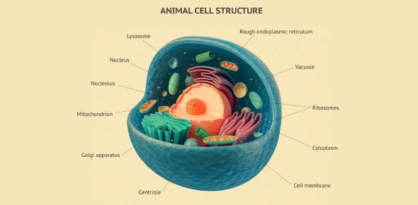
Every cell, whether it is an animal cell, plant cell, or any other type of eukaryotic cell, is composed of three fundamental components: the plasma membrane, the cytoplasm, and the nucleus. These three parts work together to maintain the cell's structure, regulate its internal environment, and control its activities.

1. Plasma Membrane

The plasma membrane, also known as the cell membrane, is the outer boundary of the cell. It is a flexible, semi-permeable membrane that encloses the cell's contents and separates it from the external environment. The plasma membrane is primarily composed of a phospholipid bilayer interspersed with proteins, cholesterol, and carbohydrates. This structure allows the membrane to be fluid and dynamic, enabling the cell to interact with its surroundings.

  • Structure
    The phospholipid bilayer consists of hydrophilic (water-attracting) heads and hydrophobic (water-repelling) tails. The hydrophilic heads face outward, toward the aqueous environments inside and outside the cell, while the hydrophobic tails face inward, away from water. Embedded within this bilayer are various proteins that serve as channels, receptors, and transporters, as well as cholesterol molecules that help maintain membrane fluidity and stability.
  • Function
    The plasma membrane controls the movement of substances into and out of the cell, maintaining the appropriate balance of ions, nutrients, and waste products. It acts as a selective barrier, allowing only certain molecules to pass through while blocking others. This selective permeability is crucial for maintaining the cell's internal environment, known as homeostasis. The membrane also plays a key role in cell communication and signaling. Receptor proteins on the surface of the membrane detect and respond to external signals, such as hormones and neurotransmitters, which can trigger specific cellular responses.

2. Cytoplasm

The cytoplasm is the jelly-like substance that fills the interior of the cell. It is composed of water, salts, and various organic molecules, including enzymes, proteins, and lipids. The cytoplasm houses all the cell's organelles, excluding the nucleus, and provides a medium for chemical reactions to occur.

  • Structure
    The cytoplasm is divided into two main parts: the cytosol and the organelles. The cytosol is the fluid component, consisting primarily of water, in which various molecules and ions are dissolved. The organelles are specialized structures within the cytoplasm that perform specific functions essential to the cell's survival and operation.
  • Function
    The cytoplasm serves as the site for many of the cell's metabolic activities. It provides a medium for biochemical reactions, such as glycolysis (the breakdown of glucose to produce energy) and protein synthesis. The cytoplasm also plays a role in intracellular transport, as molecules and organelles move through the cytoplasm to reach different parts of the cell. Additionally, the cytoplasm helps maintain the cell's shape and provides structural support for the organelles, enabling them to function efficiently.

3. Nucleus

The nucleus is the control center of the cell, responsible for storing and protecting the cell's genetic material (DNA). It regulates all cellular activities, including growth, metabolism, and reproduction, by controlling the expression of genes.

  • Structure
    The nucleus is surrounded by a double membrane called the nuclear envelope, which contains nuclear pores that allow the exchange of materials between the nucleus and the cytoplasm. Inside the nucleus, the DNA is organized into chromatin, a complex of DNA and proteins. During cell division, the chromatin condenses to form visible chromosomes. The nucleus also contains a dense, spherical structure called the nucleolus, where ribosomal RNA (rRNA) is synthesized and assembled with proteins to form ribosomes.
  • Function
    The nucleus controls the cell's activities by regulating gene expression. It does this by transcribing DNA into messenger RNA (mRNA), which is then transported out of the nucleus and translated into proteins by ribosomes in the cytoplasm. These proteins carry out various functions within the cell, such as catalyzing reactions, providing structural support, and regulating cell processes. The nucleus also plays a crucial role in cell division, ensuring that genetic material is accurately replicated and distributed to daughter cells.

Together, the plasma membrane, cytoplasm, and nucleus form the core of the cell, each playing a vital role in maintaining the cell's structure, function, and survival. These components work in concert to ensure that the cell operates efficiently, responds to environmental changes, and contributes to the organism's overall health and function.

What Are the Differences Between an Animal and Plant Cell?

Fig: Cell Anatomy of Plant and Animal Composition

Animal and plant cells, while both eukaryotic and share many common features, have several key differences that reflect their distinct roles and adaptations in living organisms. These differences can be observed in the presence or absence of certain structures, the way they produce and store energy, and their overall shape and function.

1. Cell Wall vs. Plasma Membrane

  • Plant Cells
    One of the most notable differences between plant and animal cells is the presence of a rigid cell wall in plant cells. The cell wall, primarily composed of cellulose, provides structural support and protection, allowing plants to maintain their shape and resist external forces. The cell wall also helps plants stand upright and grow tall, contributing to the overall rigidity and strength of the plant structure.
  • Animal Cells
    In contrast, animal cells lack a cell wall and are instead surrounded only by a flexible plasma membrane. This flexibility allows animal cells to adopt a variety of shapes, which is essential for the diverse functions they perform. For example, the flexibility of the plasma membrane enables animal cells to move, change shape, and form different tissues, such as muscle and nerve tissue.

2. Chloroplasts and Photosynthesis

  • Plant Cells
    Plant cells contain chloroplasts, specialized organelles that conduct photosynthesis. Chloroplasts contain chlorophyll, the green pigment that captures light energy and converts it into chemical energy in the form of glucose. This process not only provides energy for the plant's growth and development but also produces oxygen as a byproduct, which is essential for the survival of aerobic organisms, including animals.
  • Animal Cells
    Animal cells do not have chloroplasts because they do not perform photosynthesis. Instead, animal cells obtain energy by consuming organic material through processes such as cellular respiration, which occurs in the mitochondria. This difference highlights the autotrophic nature of plants (producing their own food) versus the heterotrophic nature of animals (relying on external food sources).

3. Vacuoles and Storage

  • Plant Cells
    Plant cells typically contain a large central vacuole that occupies a significant portion of the cell's volume. The central vacuole serves multiple functions, including storing water, nutrients, and waste products. It also helps maintain turgor pressure, which is essential for keeping the plant upright and supporting its structure.
  • Animal Cells
    Animal cells, on the other hand, have smaller vacuoles, and these are usually more numerous. These smaller vacuoles are primarily involved in storage and transport within the cell, but they do not play as significant a role in maintaining cell structure as the central vacuole does in plant cells.

4. Shape and Structural Organization

  • Plant Cells
    Plant cells generally have a more regular, rectangular shape due to the rigidity provided by the cell wall. This uniformity allows plant cells to form tightly packed tissues, such as the layers found in leaves, stems, and roots. The rigid structure also aids in the plant's overall stability and strength.
  • Animal Cells
    Animal cells are more varied in shape, ranging from spherical to elongated or irregular. This variety is due to the lack of a cell wall, which allows animal cells to be more flexible and dynamic in their shapes. The adaptability of animal cells is crucial for the formation of specialized tissues, such as muscles that can contract and relax, or neurons that can extend long distances to transmit signals.

5. Energy Production and Storage

  • Plant Cells
    In addition to producing energy through photosynthesis, plant cells store energy primarily in the form of starch, a polysaccharide that can be broken down into glucose when needed. This storage method is suited to the plant's need to have a readily available energy source during periods when photosynthesis is not possible, such as during the night or in low-light conditions.
  • Animal Cells
    Animal cells produce energy through cellular respiration, which occurs in the mitochondria. They store energy mainly in the form of glycogen, a highly branched polysaccharide that can be quickly mobilized to meet immediate energy needs. Additionally, animal cells store energy in the form of fats (lipids) within adipose tissue, which provides a long-term energy reserve.

What Is the Function of the Animal Cell?

The primary function of an animal cell is to perform the various processes necessary for the survival, growth, and overall function of the organism. Each animal cell operates as a self-contained unit, carrying out essential tasks that contribute to the organism's health and functionality. These functions include energy production, protein synthesis, waste management, and cellular regulation, all of which are performed by the cell's organelles working in concert.

  1. Energy Production
    • The mitochondria, often referred to as the "powerhouses" of the cell, are responsible for generating energy. Through the process of cellular respiration, mitochondria convert glucose and oxygen into adenosine triphosphate (ATP), the primary energy currency of the cell. This energy is used to power various cellular activities, from muscle contraction to cell division. Without sufficient energy production, the cell would be unable to sustain its vital functions, leading to cell death and, consequently, harm to the organism.
  2. Protein Synthesis
    • Protein synthesis is a critical function of the cell, as proteins play a multitude of roles within the body, including acting as enzymes, structural components, and signaling molecules. Ribosomes, either floating freely in the cytoplasm or attached to the rough endoplasmic reticulum (ER), are the sites of protein synthesis. These organelles read the genetic instructions encoded in messenger RNA (mRNA) and assemble amino acids into specific sequences to form proteins. The proteins synthesized by ribosomes are then folded, modified, and transported to their appropriate destinations within or outside the cell, where they perform their designated functions.
  3. Regulation of Cellular Activities
    • The nucleus, which contains the cell's genetic material (DNA), acts as the control center of the cell. It regulates all cellular activities by controlling gene expression and coordinating cellular responses to internal and external signals. The nucleus ensures that the cell functions in harmony with other cells in the body, maintaining homeostasis and supporting the organism's overall health. Through processes such as DNA replication, transcription, and mRNA processing, the nucleus directs the production of proteins and other molecules that are essential for the cell's operation.
  4. Cellular Communication and Interaction
    • Animal cells must communicate with each other and their environment to coordinate activities and maintain the organism's overall function. The plasma membrane plays a crucial role in this communication by hosting various receptors and channels that detect signals from other cells, such as hormones or neurotransmitters. These signals are then transmitted into the cell, triggering specific responses that can affect the cell's behavior, such as altering its metabolic activity, initiating cell division, or moving toward a signal source.
  5. Waste Management and Recycling
    • The lysosomes, often described as the cell's waste disposal system, are responsible for breaking down and recycling cellular waste and debris. These organelles contain powerful digestive enzymes that degrade damaged organelles, macromolecules, and pathogens that enter the cell. By efficiently managing waste, lysosomes help prevent the accumulation of toxic substances within the cell, ensuring that the cellular environment remains healthy and conducive to proper functioning.
  6. Structural Integrity and Support
    • The cytoskeleton, a network of protein fibers that includes microtubules, microfilaments, and intermediate filaments, provides structural support to the cell. It helps maintain the cell's shape, anchors organelles in place, and facilitates cellular movement and division. The cytoskeleton also plays a role in intracellular transport, guiding vesicles and organelles to their correct locations within the cell. This structural framework is essential for the cell's mechanical stability and its ability to interact dynamically with its environment. In summary, the primary function of an animal cell is to coordinate and execute a wide range of processes that are essential for the survival and function of the organism. Each organelle within the cell contributes to these processes, ensuring that the cell operates efficiently and supports the larger organism.

What Are Cell Structures and Their Functions?

Fig: Structure of an Animal Cell

Animal cells are composed of various specialized structures, known as organelles, each of which has specific functions that contribute to the cell's overall operation. These organelles work together to ensure that the cell remains healthy, performs its necessary tasks, and contributes to the organism's overall vitality. Below is a detailed overview of key cell structures and their functions:

  1. Nucleus
    • The nucleus serves as the control center of the cell. It houses the cell's genetic material (DNA), which contains the instructions for all cellular activities. The nucleus regulates processes such as gene expression, cell growth, and replication by controlling the transcription of DNA into messenger RNA (mRNA). The mRNA is then used to synthesize proteins, which are essential for the cell's functions. The nucleus is surrounded by a double membrane called the nuclear envelope, which contains nuclear pores that allow the exchange of materials between the nucleus and the cytoplasm.
  2. Mitochondria
    • Mitochondria are the energy-generating organelles of the cell, often referred to as the "powerhouses" of the cell. They produce ATP through the process of cellular respiration, which involves the breakdown of glucose and other nutrients in the presence of oxygen. The energy produced by mitochondria is used to power various cellular processes, including muscle contraction, cell division, and the synthesis of macromolecules. Mitochondria have their own DNA and are believed to have originated from ancient symbiotic bacteria.
  3. Ribosomes
    • Ribosomes are the molecular machines responsible for protein synthesis. They read the genetic instructions carried by mRNA and assemble amino acids into polypeptide chains, which fold into functional proteins. Ribosomes can be found floating freely in the cytoplasm or attached to the rough endoplasmic reticulum (ER). The proteins synthesized by ribosomes play crucial roles in the cell, including serving as enzymes, structural components, and signaling molecules.
  4. Endoplasmic Reticulum (ER)
    • The endoplasmic reticulum is a network of membranous tubules and sacs that plays a key role in the synthesis, folding, modification, and transport of proteins and lipids. There are two types of ER
      • Rough ER
        Studded with ribosomes, the rough ER is primarily involved in the synthesis and processing of proteins destined for secretion, incorporation into the cell membrane, or use in lysosomes.
      • Smooth ER
        Lacking ribosomes, the smooth ER is involved in the synthesis of lipids, metabolism of carbohydrates, detoxification of drugs and poisons, and storage of calcium ions. The smooth ER also plays a role in the production of steroid hormones.
  5. Golgi Apparatus
    • The Golgi apparatus, also known as the Golgi complex or Golgi body, is a series of flattened, membrane-bound sacs that modify, sort, and package proteins and lipids for transport to their final destinations within or outside the cell. The Golgi apparatus is involved in the processing of proteins synthesized in the rough ER, adding carbohydrates to form glycoproteins, and packaging them into vesicles for delivery. The Golgi apparatus also plays a role in the formation of lysosomes.
  6. Lysosomes
    • Lysosomes are membrane-bound organelles that contain digestive enzymes capable of breaking down macromolecules, damaged organelles, and pathogens. They play a crucial role in cellular waste disposal and recycling, ensuring that the cell remains free of debris and harmful substances. Lysosomes are also involved in processes such as autophagy, where the cell degrades and recycles its own components to maintain cellular health and homeostasis.
  7. Cytoskeleton
    • The cytoskeleton is a dynamic network of protein fibers that provides structural support to the cell, helps maintain its shape, and facilitates movement. The cytoskeleton is composed of three main types of fibers
      • Microtubules
        Hollow tubes that provide a framework for organelle movement, cell division, and the formation of cilia and flagella.
      • Microfilaments
        Thin, thread-like structures made of actin that play a role in cell movement, muscle contraction, and cell division.
      • Intermediate Filaments
        Rope-like fibers that provide tensile strength, helping to maintain cell shape and anchor organelles in place.

These organelles work together to perform the complex tasks necessary for the cell's survival, growth, and function. By coordinating their activities, the organelles ensure that the cell operates efficiently and contributes to the overall health and vitality of the organism.

What Are the Features of an Animal Cell?

Animal cells possess several distinct features that differentiate them from plant cells and other cell types. These features are essential for the specialized functions that animal cells perform within the body. Understanding these features helps to clarify the unique roles that animal cells play in maintaining the health and functionality of the organism.

  1. Flexible Plasma Membrane
    • Unlike plant cells, animal cells do not have a rigid cell wall. Instead, they are surrounded by a flexible plasma membrane that allows them to adopt various shapes and forms. This flexibility is crucial for the dynamic activities that animal cells perform, such as movement, communication, and the formation of tissues. The plasma membrane also plays a key role in regulating the exchange of substances between the cell and its environment, maintaining homeostasis.
  2. Presence of Lysosomes
    • Lysosomes are a characteristic feature of animal cells, involved in the digestion and recycling of cellular waste. These organelles contain hydrolytic enzymes that break down macromolecules, damaged organelles, and pathogens. The presence of lysosomes ensures that the cell remains clean and free of debris, which is essential for maintaining cellular health and preventing the buildup of harmful substances.
  3. Absence of a Cell Wall and Chloroplasts
    • One of the most significant differences between animal and plant cells is the absence of a cell wall in animal cells. The lack of a cell wall allows animal cells to be more flexible and adaptable in their shapes, enabling them to perform a wide range of functions, such as forming different types of tissues and responding to environmental changes. Additionally, animal cells do not contain chloroplasts, as they do not perform photosynthesis. Instead, animal cells rely on mitochondria to produce energy through cellular respiration, using organic molecules obtained from food.
  4. Varied Shapes and Sizes
    • Animal cells exhibit a wide variety of shapes and sizes, reflecting their specialized functions within the body. For example, nerve cells (neurons) have long extensions that enable them to transmit signals over long distances, while red blood cells are small and flexible, allowing them to navigate through narrow blood vessels. Muscle cells are elongated and packed with contractile proteins, enabling them to generate force and movement. This diversity in form allows animal cells to perform the complex and varied functions required for the organism's survival.
  5. Dynamic Cytoskeleton
    • The cytoskeleton is a defining feature of animal cells, providing structural support and enabling movement. The cytoskeleton is composed of microtubules, microfilaments, and intermediate filaments, each of which plays a role in maintaining cell shape, facilitating intracellular transport, and driving cell division. The dynamic nature of the cytoskeleton allows animal cells to adapt to changing conditions, move in response to signals, and organize their internal components efficiently.

What Are the Different Types of Animal Cells?

Fig: Different Types of Cells in Humans

Animal cells exhibit a remarkable diversity in structure and function, reflecting the wide range of roles they play within the body. This diversity allows for the specialization of cells to perform specific tasks necessary for the survival, growth, and health of the organism. Below is an exploration of some of the most common types of animal cells, categorized based on their structure and function.

1. Epithelial Cells

Epithelial cells form the protective covering or lining of organs, cavities, and surfaces throughout the body. They are tightly packed together, creating barriers that protect underlying tissues from mechanical damage, pathogens, and dehydration.

  • Structure
    Epithelial cells are generally polygonal in shape and can be classified based on the number of layers they form and the shape of the cells themselves. Simple epithelium consists of a single layer of cells, while stratified epithelium has multiple layers. The cells can be squamous (flat), cuboidal (cube-shaped), or columnar (tall and cylindrical).
  • Function
    Epithelial cells serve several critical functions, including protection, absorption, secretion, and sensation. For example, the epithelial cells lining the intestines absorb nutrients from digested food. Those in the respiratory tract secrete mucus to trap and remove dust and pathogens. In the skin, epithelial cells form a barrier that protects against environmental damage and water loss. Additionally, some epithelial cells have specialized structures, such as cilia, that help move substances across the cell surface.

2. Muscle Cells

Muscle cells, also known as myocytes, are specialized for contraction and movement. They are responsible for generating force and movement, enabling activities such as locomotion, posture maintenance, and the pumping of blood through the heart.

  • Structure
    Muscle cells are elongated and packed with contractile proteins, primarily actin and myosin, which slide past each other to produce contraction. Muscle cells can be categorized into three types: skeletal, cardiac, and smooth.
    • Skeletal Muscle Cells
      Skeletal muscle cells are long, cylindrical, and multinucleated, with a striated appearance due to the regular arrangement of actin and myosin filaments. These cells are under voluntary control and are attached to bones, enabling body movements.
    • Cardiac Muscle Cells
      Cardiac muscle cells are found only in the heart. They are branched and interconnected, forming a network that allows the heart to contract in a coordinated manner. These cells have a single nucleus and are also striated. They are under involuntary control and are responsible for pumping blood throughout the body.
    • Smooth Muscle Cells
      Smooth muscle cells are spindle-shaped, with a single central nucleus, and lack striations. These cells are found in the walls of hollow organs, such as the intestines, blood vessels, and bladder. They are under involuntary control and help regulate internal processes, such as the movement of food through the digestive tract and the regulation of blood flow.
  • Function
    The primary function of muscle cells is to generate force and produce movement. Skeletal muscle cells enable voluntary movements, such as walking and lifting objects. Cardiac muscle cells maintain the rhythmic contractions of the heart, ensuring continuous blood circulation. Smooth muscle cells control involuntary movements, such as the constriction and relaxation of blood vessels and the peristaltic movement of the digestive tract.

3. Nerve Cells (Neurons)

Nerve cells, or neurons, are specialized for communication and signal transmission. They form the core components of the nervous system, which includes the brain, spinal cord, and peripheral nerves.

  • Structure
    Neurons are characterized by their unique shape, which includes a cell body (soma), dendrites, and an axon.
    • Cell Body (Soma)
      The cell body contains the nucleus and most of the cell's organelles. It is responsible for maintaining the neuron's health and integrating incoming signals.
    • Dendrites
      Dendrites are branched extensions from the cell body that receive signals from other neurons or sensory receptors. They transmit these signals toward the cell body.
    • Axon
      The axon is a long, slender projection that carries electrical impulses away from the cell body toward other neurons, muscles, or glands. The axon is often insulated by a myelin sheath, which increases the speed of signal transmission.
  • Function
    Neurons are responsible for transmitting electrical and chemical signals throughout the body, enabling communication between different parts of the nervous system. This allows for the coordination of complex processes such as movement, sensation, cognition, and autonomic functions like heart rate and digestion. Neurons play a crucial role in reflexes, sensory perception, and the processing of information in the brain.

4. Blood Cells

Blood cells are specialized cells that circulate within the bloodstream, performing a variety of critical functions, including oxygen transport, immune response, and clotting.

  • Structure
    Blood cells can be broadly categorized into three main types: red blood cells (erythrocytes), white blood cells (leukocytes), and platelets (thrombocytes).
    • Red Blood Cells (Erythrocytes)
      Red blood cells are biconcave and lack a nucleus, which increases their surface area for oxygen transport. They contain the protein hemoglobin, which binds to oxygen in the lungs and releases it in tissues throughout the body.
    • White Blood Cells (Leukocytes)
      White blood cells are a diverse group of cells involved in the immune response. They include lymphocytes (such as T cells and B cells), monocytes, neutrophils, eosinophils, and basophils. Each type of white blood cell has a specific role in defending the body against infections, foreign invaders, and abnormal cells.
    • Platelets (Thrombocytes)
      Platelets are small, cell-fragment-like structures that play a key role in blood clotting. They adhere to damaged blood vessels and release factors that initiate the clotting process, preventing excessive blood loss during injury.
  • Function
    Blood cells perform a variety of essential functions that are crucial for maintaining the body's homeostasis.
    • Red Blood Cells
      Red blood cells transport oxygen from the lungs to tissues and carry carbon dioxide from tissues back to the lungs for exhalation.
    • White Blood Cells
      White blood cells protect the body against infections and foreign invaders by identifying, attacking, and destroying pathogens and abnormal cells.
    • Platelets
      Platelets participate in the formation of blood clots to prevent bleeding and facilitate wound healing.

5. Connective Tissue Cells

Connective tissue cells provide structural support, connect different tissues, and protect organs. They are an integral part of the body's framework and play a role in tissue repair and the storage of energy.

  • Structure
    Connective tissue cells include a variety of cell types, such as fibroblasts, adipocytes, chondrocytes, and osteocytes.
    • Fibroblasts
      Fibroblasts are spindle-shaped cells that produce and secrete extracellular matrix components, such as collagen and elastin, which provide strength and elasticity to tissues.
    • Adipocytes
      Adipocytes, or fat cells, are specialized for storing energy in the form of lipids. They have a large central lipid droplet that occupies most of the cell's volume.
    • Chondrocytes
      Chondrocytes are the cells found in cartilage, where they produce and maintain the cartilaginous matrix, which provides cushioning and flexibility to joints.
    • Osteocytes
      Osteocytes are mature bone cells that maintain the mineralized bone matrix. They are involved in bone remodeling and the regulation of calcium and phosphate levels.
  • Function
    Connective tissue cells perform several important functions within the body.
    • Fibroblasts
      Fibroblasts provide structural support by producing extracellular matrix components, which form the framework for tissues and organs.
    • Adipocytes
      Adipocytes store energy in the form of fat and release it when needed, playing a role in energy balance and insulation.
    • Chondrocytes
      Chondrocytes maintain cartilage, which provides smooth surfaces for joint movement and absorbs shock.
    • Osteocytes
      Osteocytes maintain bone tissue, support the skeleton, and regulate mineral homeostasis.

Take These Quizzes

Conclusion

In conclusion, our exploration of animal cells has provided a detailed understanding of the essential roles these cells play in the animal kingdom. We have examined the unique structures and functions of animal cells, such as the plasma membrane, nucleus, and mitochondria, which are crucial for energy production, protein synthesis, and overall cellular regulation. The diversity of animal cells, from epithelial cells to neurons, highlights their specialized functions that support the organism's health and survival.

Additionally, by tracing the historical development of cell theory, we have appreciated the foundational principles that underpin modern biology. Understanding the differences between animal and plant cells further enriches our comprehension of how these cells contribute to life's complexity. The study of animal cells continues to be central to advancements in understanding life and improving health outcomes.

Back to Top Back to top
Advertisement
×

Wait!
Here's an interesting quiz for you.

We have other quizzes matching your interest.