Apoptosis Definition, Steps, Significance & Role: A Complete Guide with Real-World Relevance
Lesson Overview
This lesson on apoptosis covers its definition with steps, its significance, and role in biological processes. Understanding apoptosis is essential for studying cell regulation, disease prevention, and developmental biology. By learning this topic, you will gain insight into how controlled cell death maintains health and stability in multicellular organisms.
What Is Apoptosis?
Apoptosis is a programmed process where cells self-destruct in a controlled manner to maintain tissue health. It prevents damaged, unnecessary, or harmful cells from accumulating. Unlike necrosis, which is uncontrolled cell death due to injury, apoptosis is regulated and beneficial.
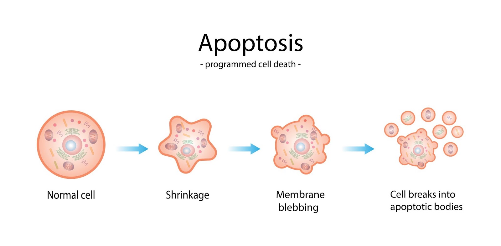
Fig: A diagram of apoptosis showing a cell undergoing programmed cell death in a controlled manner, compared to necrosis, which results from injury.
Apoptosis Examples:
- During fetal development, apoptosis shapes fingers and toes by removing webbing.
- In the immune system, defective or infected cells undergo apoptosis to prevent disease.
- Damaged skin cells exposed to UV radiation die via apoptosis to prevent cancer.
Why Does Apoptosis Happen?
Apoptosis happens when a cell receives internal or external signals that activate a controlled cell death process. The process is triggered by various factors, including DNA damage, lack of survival signals, or immune system regulation.
- Intrinsic Pathway (Mitochondrial Pathway)
- When a cell experiences stress (e.g., DNA damage, oxidative stress, or lack of nutrients), proteins like p53 detect the damage.
- The mitochondria release cytochrome c, which binds to Apaf-1, forming the apoptosome.
- The apoptosome activates caspase-9, which then activates executioner caspases (caspase-3, caspase-7) to break down the cell.
- Extrinsic Pathway (Death Receptor Pathway)
- External signals, such as immune system proteins (TNF-α, Fas ligand), bind to death receptors (Fas, TNFR) on the cell membrane.
- These receptors activate caspase-8, which directly activates executioner caspases.
- Executioner caspases degrade proteins and DNA, leading to controlled cell fragmentation.
- Cellular Breakdown
- The cytoskeleton collapses, and the nucleus condenses (pyknosis).
- The DNA is broken into small fragments (karyorrhexis).
- The cell membrane forms small vesicles called apoptotic bodies, which are engulfed by phagocytes without causing inflammation.
Take This Quiz :
(102).jpg)
What Is the Function of Apoptosis?
The purpose of apoptosis is to eliminate unnecessary, damaged, or potentially harmful cells in a controlled manner. This prevents diseases, maintains homeostasis, and supports proper development.
1. Maintaining Tissue Homeostasis
- The body constantly produces new cells, and apoptosis removes old or unneeded ones to maintain a healthy balance.
- In organs like the skin and intestines, apoptosis ensures that dead cells are replaced without excessive tissue buildup.
2. Supporting Embryonic Development
- During fetal development, apoptosis shapes body structures by removing unwanted cells.
- Example: Finger and toe formation occurs when apoptosis eliminates the webbing between digits.
3. Removing Damaged or Mutated Cells
- Cells with irreversible DNA damage due to radiation, toxins, or replication errors undergo apoptosis to prevent mutations from spreading.
- This acts as a natural defense mechanism against cancer, ensuring that defective cells do not continue dividing.
4. Regulating the Immune System
- Apoptosis eliminates excess immune cells after an infection is cleared, preventing unnecessary inflammation.
- It also removes self-reactive immune cells that could attack the body's tissues, reducing the risk of autoimmune diseases.
5. Preventing Uncontrolled Cell Growth (Cancer Prevention)
- When cells divide uncontrollably due to faulty apoptosis mechanisms, tumors can form.
- The purpose of apoptosis is to act as a built-in safeguard, destroying potentially cancerous cells before they proliferate.
6. Removing Infected or Harmful Cells
- Cells infected with viruses often undergo apoptosis to prevent the spread of infection.
- This is a key defense strategy in the immune system, helping the body fight off diseases.
Key Molecules Involved in Apoptosis
Apoptosis is regulated by a network of proteins that control cell death in a precise manner. The key molecules involved in apoptosis include:
1. Caspases (Executioner Proteins)
- Caspases are a family of proteolytic enzymes that act as the main executioners of apoptosis.
- They exist as inactive precursors (procaspases) and get activated in response to apoptotic signals.
- Key caspases:
- Initiator caspases (Caspase-8, Caspase-9) – Trigger apoptosis.
- Executioner caspases (Caspase-3, Caspase-6, Caspase-7) – Break down cellular components.
2. Bcl-2 Family Proteins (Pro- and Anti-Apoptotic Regulators)
- The Bcl-2 protein family controls the mitochondrial pathway of apoptosis.
- It includes pro-apoptotic proteins (Bax, Bak) and anti-apoptotic proteins (Bcl-2, Bcl-xL).
- Balance between these proteins determines whether a cell survives or undergoes apoptosis.
3. Cytochrome c (Mitochondrial Death Signal)
- It is released from mitochondria when the apoptotic pathway is activated.
- It binds with Apaf-1 (Apoptotic Protease Activating Factor-1) to form the apoptosome, which activates caspases.
4. Death Receptors (Extrinsic Pathway Triggers)
- It is present on the cell surface, these receptors activate apoptosis when bound by specific ligands.
- Examples:
- Fas receptor (CD95) – Activated by Fas ligand (FasL).
- TNF receptor (TNFR1) – Activated by Tumor Necrosis Factor (TNF-α).
5. p53 (Guardian of the Genome)
- A tumor suppressor protein that detects DNA damage and triggers apoptosis if the damage is beyond repair.
- Prevents cancer development by eliminating potentially cancerous cells.
Stages of Apoptosis
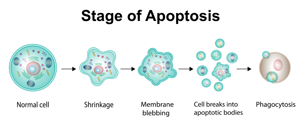
Apoptosis occurs in a series of controlled steps to ensure the orderly removal of dying cells. The steps of apoptosis can be divided into the following stages:
1. Initiation Stage
- The process begins when a cell receives an apoptotic signal.
- Apoptosis can be triggered by two pathways:
- Intrinsic pathway (mitochondrial-mediated, triggered by DNA damage or stress).
- Extrinsic pathway (death receptor-mediated, triggered by external signals like FasL or TNF-α).
2. Activation of Caspases
- Initiator caspases (Caspase-8, Caspase-9) get activated.
- These activate executioner caspases (Caspase-3, Caspase-6, Caspase-7), which begin breaking down cellular components.
3. Cell Shrinkage and Chromatin Condensation
- The cell becomes smaller due to cytoskeleton breakdown.
- The nucleus condenses (pyknosis), and DNA is fragmented (karyorrhexis).
4. Membrane Blebbing and Organelle Breakdown
- The plasma membrane forms small protrusions (blebs).
- The Golgi apparatus, endoplasmic reticulum, and mitochondria break down in a controlled manner.
5. Formation of Apoptotic Bodies
- The cell fragments into membrane-bound apoptotic bodies containing cellular debris.
- These are quickly recognized and cleared by immune cells.
6. Phagocytosis of Apoptotic Bodies
- Macrophages or neighboring cells engulf and digest apoptotic bodies without triggering inflammation.
Take This Quiz :

Why Is Apoptosis Important?
Let's understand it simply:
- It stops damaged cells from growing into tumors.
- It removes infected cells to keep the immune system healthy.
- It helps shape organs in a growing baby.
- It replaces old cells to keep tissues healthy.
- It stops the immune system from attacking the body.
- It clears extra brain cells to keep nerves working properly.
What Is the End Result of Apoptosis?
The end result of apoptosis is the controlled elimination of unwanted, damaged, or aged cells without causing harm to surrounding tissues. This ensures proper development, immune function, and tissue homeostasis.
- Cellular Breakdown into Apoptotic Bodies
- The dying cell fragments into small, membrane-bound vesicles called apoptotic bodies that contain cellular components.
- Phagocytosis by Immune or Neighboring Cells
- Macrophages or nearby cells engulf and digest apoptotic bodies, preventing inflammation and tissue damage.
- No Release of Harmful Substances
- Unlike necrosis, apoptosis does not cause inflammation because cell contents remain enclosed within apoptotic bodies.
- Tissue Remodeling and Homeostasis
- Apoptosis helps in shaping organs during development (e.g., removal of webbing between fingers in embryos).
- It maintains healthy cell turnover in tissues like the skin, gut lining, and immune system.
- Prevention of Disease
- Proper apoptosis removes potentially cancerous or virus-infected cells, reducing the risk of disease.
- Failure of apoptosis can lead to disorders like cancer, autoimmune diseases, or neurodegeneration.
Rate this lesson:
 Back to top