Staining in Microbiology: Definitions, Types, and Core Lab Techniques
Lesson Overview
Staining is a way to make tiny things, like cells, easier to see under a microscope. Most cells are clear, so without staining, it's hard to tell what's inside them. Stains are like special dyes that color parts of the cells, helping us see their shapes and structures clearly.
For example, staining can show us the nucleus of a cell or help us tell the difference between two types of bacteria. This simple technique is used in science labs to study plants, animals, and even diseases. By learning about staining, you'll understand how scientists explore the microscopic world with greater detail and precision.
What Is Staining?
Staining is a laboratory technique used to add color to cells, tissues, or microorganisms, making them easier to observe under a microscope. It involves applying specific dyes that bind to different parts of the specimen, highlighting their structures.
This method helps distinguish components like the cell wall, nucleus, or cytoplasm, allowing scientists to study their shapes, sizes, and functions. Staining is essential in identifying microorganisms, analyzing cell components, and diagnosing diseases.
Take This Quiz :

Types of Staining Techniques
Staining techniques vary depending on the purpose of the observation and the structures being studied. Each method uses specific dyes to highlight unique features of cells, tissues, or microorganisms, providing crucial insights for research and diagnostics.
Gram Staining:
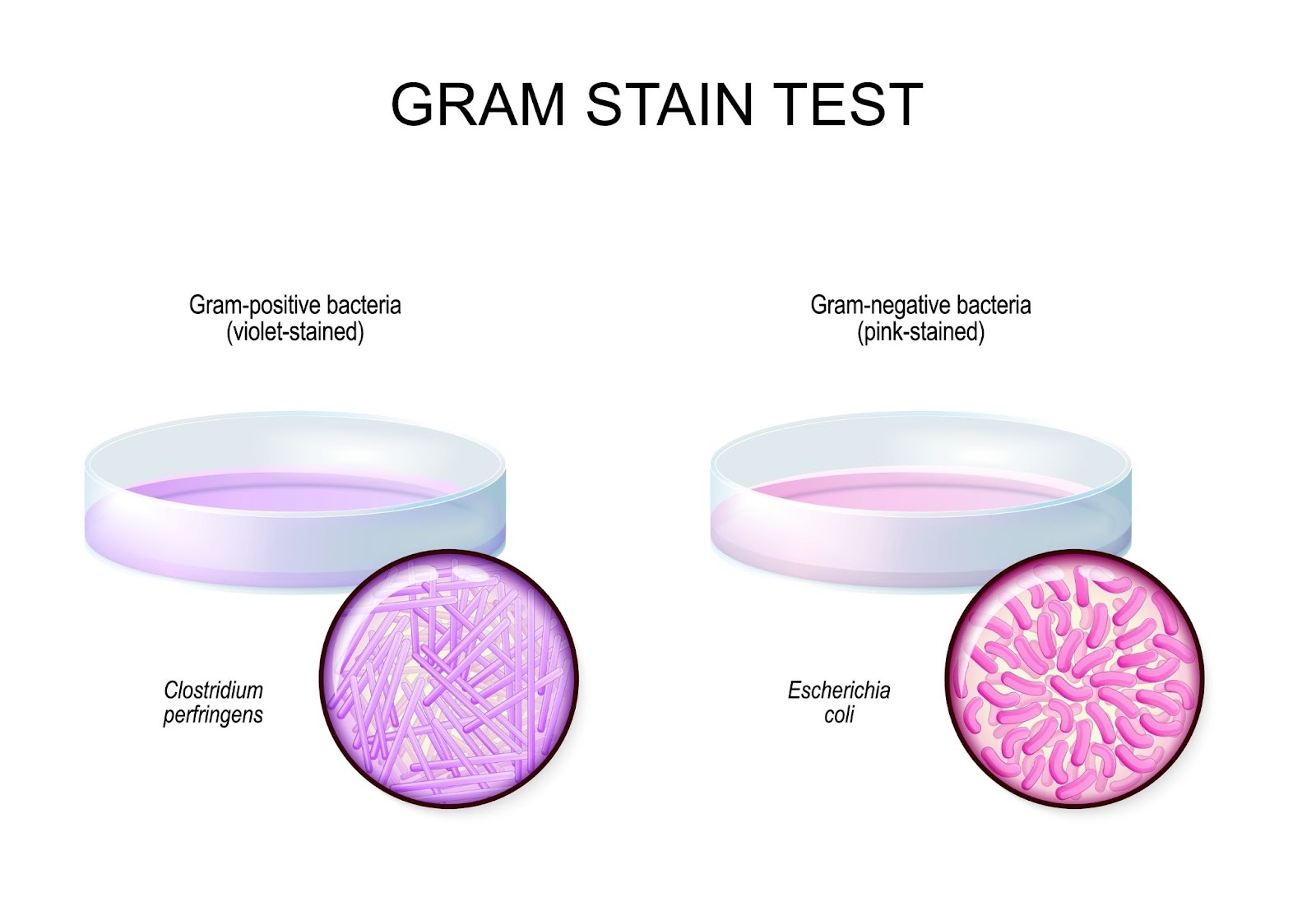
Gram staining is a key technique to classify bacteria based on their cell wall structure into Gram-positive or Gram-negative. It uses crystal violet as the primary stain, iodine as a mordant, alcohol for decolorization, and safranin as a counterstain. This method helps identify bacterial types, aiding in diagnosis and treatment.
Example: Escherichia coli (Gram-negative) and Clostridium perfringens (Gram-positive) are common examples that can be demonstrated to show the difference in cell wall composition.
Endospore Staining:
Endospore staining is used to detect bacterial endospores, which are resistant, dormant structures. The process involves heat to help malachite green penetrate the spore, followed by safranin to stain the surrounding cells. Endospores are critical to study because they make bacteria harder to eliminate.
Example: Bacillus subtilis or Clostridium difficile can be used to show the presence of endospores and how they resist staining without special techniques.
Acid-Fast Staining (Ziehl-Neelsen):
This technique targets bacteria with waxy cell walls, like Mycobacterium tuberculosis. It uses carbol fuchsin to stain the bacteria and methylene blue as a counterstain. Acid-fast staining is essential for identifying pathogens that don't respond to Gram staining.
Example: Mycobacterium tuberculosis or Mycobacterium leprae are ideal examples to show how acid-fast bacteria resist Gram staining and require this specialized technique.
Hematoxylin and Eosin (H&E) Staining:
Commonly used in histology, this method highlights tissue structures. Hematoxylin stains nuclei blue, while eosin stains the cytoplasm and extracellular components pink or red. It's widely applied in medical pathology for tissue examination.
Example: Sections of human liver tissue or kidney tissue can be used to observe nuclei and cytoplasm staining, helping students understand its role in histology.
Periodic Acid-Schiff (PAS) Staining:
PAS staining identifies carbohydrates like glycogen in tissues. Periodic acid oxidizes carbohydrates, allowing Schiff reagent to bind and produce a magenta color. It's often used to study liver, kidney, and other tissues for disease diagnosis.
Example: Staining a section of liver tissue with glycogen deposits will demonstrate how PAS staining highlights carbohydrates.
Masson's Trichrome Staining:
This three-color staining technique differentiates cells from connective tissues. It stains muscle fibers and keratin red, collagen blue or green, and nuclei black. It is commonly applied in research involving muscle and connective tissue structure.
Example: A muscle biopsy or connective tissue sample can be used to show the differentiation between muscle fibers (red) and collagen (blue or green).
Take This Quiz :
(187).webp)
How Does Staining Work?
Staining is a key technique in microscopy that helps make invisible details visible. It works by using chemical dyes to highlight specific parts of cells or tissues, allowing scientists to observe their structure and function clearly.
- Interaction with Specimen:
Stains are chemical dyes designed to react with specific cellular components. When applied, they chemically bind to molecules like proteins, lipids, carbohydrates, or nucleic acids, forming a visible color contrast. This binding is crucial for identifying the unique features of the specimen.
- Stain Penetration:
The dye penetrates the specimen through absorption. Heat or chemicals may be used to assist the stain in entering the cell or tissue. For example, in endospore staining, heat is applied to allow the dye to pass through the tough spore coat.
- Selective Binding:
Different stains bind to specific parts of the specimen based on their chemical properties. Basic dyes (positively charged) like crystal violet bind to negatively charged cell structures, such as nucleic acids. Conversely, acidic dyes (negatively charged) like eosin bind to positively charged proteins. This selective binding highlights different structures in unique colors.
- Contrast Formation:
Transparent specimens are difficult to observe under a microscope. Staining adds contrast by coloring particular parts of the specimen, making them stand out from the background. For instance, Gram staining differentiates bacteria into Gram-positive (purple) and Gram-negative (pink), improving the visibility of cell wall differences.
- Fixation and Counterstaining:
Fixation preserves the specimen's structure and prevents distortion during staining. Some techniques use a counterstain to provide additional contrast. For example, in Gram staining, safranin is used as a counterstain to color Gram-negative bacteria, creating a clear distinction between bacterial types.
Take This Quiz :
(204).webp)
Steps to Perform Staining in a Lab
Staining in a laboratory follows a systematic process to ensure clarity and accuracy in microscopic observation. Below are the key steps involved:
- Preparation of the Slide:
Start by placing the specimen (such as a bacterial smear or tissue sample) on a clean glass slide. If required, thin the sample to create a single layer for better staining results.
- Fixation:
Fix the specimen to the slide using heat or chemicals like ethanol. This step ensures the specimen adheres to the slide, preserves its structure, and prevents distortion during staining.
- Application of Primary Stain:
Apply the primary stain to the specimen and let it sit for a specific amount of time. This dye interacts with targeted cellular structures to highlight specific features. For instance, in Gram staining, crystal violet is the primary stain.
- Rinsing:
Gently rinse the slide with water or another solution to remove excess stain. This prevents overstaining, ensuring only the desired structures retain the color.
- Use of Mordant (if required):
Add a mordant, such as iodine in Gram staining, to fix the primary stain. The mordant strengthens the bond between the stain and the specimen, making the color more stable and vibrant.
- Decolorization (if applicable):
In differential staining methods, apply a decolorizing agent (e.g., alcohol in Gram staining) to remove the stain from certain parts of the specimen. This step creates contrast by differentiating structures that retain the primary stain from those that do not.
- Counterstaining:
Apply a secondary or counterstain to the slide. This step colors the structures that did not retain the primary stain, enhancing contrast. For example, safranin is used in Gram staining to color Gram-negative bacteria.
- Final Rinsing and Drying:
Rinse the slide gently to remove any residual dye and let it air dry or blot it carefully using filter paper.
- Microscopic Observation:
Place the prepared slide under a microscope to observe the stained specimen. Adjust the focus and lighting for a clear and detailed view.
Key Applications of Staining
Staining techniques play a crucial role in various scientific fields, enabling researchers and professionals to study microscopic structures with greater clarity. Below are the primary applications of staining:
- Microbial Identification:
Staining is widely used in microbiology to identify and classify microorganisms. Techniques like Gram staining help differentiate bacteria based on their cell wall properties, aiding in diagnosis and treatment decisions.
- Cellular and Tissue Analysis:
In biology and medicine, staining allows the detailed examination of cells and tissues. Techniques such as Hematoxylin and Eosin (H&E) staining highlight cell nuclei and cytoplasm, providing insights into cellular structure and function.
- Disease Diagnosis:
Pathologists use staining to detect abnormalities in tissues, such as cancerous cells or infections. For instance, PAS staining is used to identify glycogen storage diseases or fungal infections.
- Study of Structural Components:
Staining helps visualize specific components like proteins, lipids, or nucleic acids. For example, endospore staining identifies bacterial endospores, while Masson's trichrome distinguishes connective tissue from muscle.
- Research in Molecular Biology:
Advanced staining methods, like fluorescence staining, are used in molecular biology to study DNA, RNA, and protein interactions. This application is essential for understanding cellular processes and genetic research.
- Histopathology and Cytopathology:
In medical fields, staining techniques assist in diagnosing tissue diseases and cellular disorders. For example, Ziehl-Neelsen staining is critical for detecting tuberculosis bacteria in clinical samples.
- Observation of Living Cells:
Certain stains, like vital stains, are used to observe live cells without harming them. This is useful for studying dynamic processes like cell division and movement.
- Educational Purposes:
In educational settings, staining is used to teach students about cell structures, microbial diversity, and tissue organization, making complex biological concepts easier to understand.
Take This Quiz :
(194).webp)
Rate this lesson:
 Back to top