Mitosis Definition, Stages, Diagram, and Phases: A Complete Guide with Real-World Relevance
Life is built on growth, repair, and reproduction, and mitosis plays a key role in all three. Every second, millions of cells in your body divide to replace old, damaged, or dead cells. This process ensures that new cells carry the same genetic information as the parent cell, helping maintain the body's structure and function. Mitosis is crucial for growth in multicellular organisms-from a single fertilized egg to a fully developed human.
What Is Mitosis?
Mitosis is a process of cell division where a single cell divides to form two genetically identical daughter cells, each with the same number of chromosomes as the parent cell. This ensures the accurate transfer of genetic material for proper growth, repair, and reproduction.
Importance of Mitosis:
- Maintains Genetic Stability: Ensures each new cell has the correct chromosome number.
- Supports Growth: Helps multicellular organisms grow by increasing cell numbers.
- Aids Tissue Repair: Replaces damaged or dead cells for tissue maintenance.
- Enables Asexual Reproduction: Allows certain organisms to reproduce without genetic variation.
Where Mitosis Occurs
Mitosis occurs in specific cells and tissues that support growth, repair, and reproduction. The process takes place in various organisms and cell types depending on their biological functions.
- Somatic Cells (Body Cells):
Mitosis primarily occurs in somatic cells, which make up most of an organism's tissues and organs. Skin, liver, muscle, and bone cells undergo mitosis regularly to replace damaged, dead, or aging cells. This process maintains tissue integrity and ensures the body's proper functioning. - Asexually Reproducing Organisms:
In unicellular organisms like amoebas and bacteria, mitosis drives asexual reproduction. Through binary fission or budding, these organisms create genetically identical offspring without the need for gametes, ensuring rapid population growth. - Tissue-Specific Regions (Meristematic Tissue in Plants):
In plants, mitosis occurs in meristematic tissues found in root tips, shoot tips, and cambium layers. These regions contain actively dividing cells that support continuous growth, wound healing, and organ formation throughout the plant's life cycle. - Stem Cells in Animals:
Stem cells undergo mitosis to generate new cells for growth and tissue repair. For example, hematopoietic stem cells in bone marrow divide to produce blood cells essential for immune function and oxygen transport.
Take This Quiz :
.webp)
Features of Mitosis
Mitosis ensures the accurate division of genetic material, producing two identical daughter cells. It plays a crucial role in growth, repair, and asexual reproduction. Here are its key features:
- Occurs in Eukaryotic Cells Only
Mitosis occurs exclusively in eukaryotic cells, which have a defined nucleus and complex organelles. Prokaryotic cells, like bacteria, divide through a simpler process called binary fission.
- Involves Spindle Fiber Formation
Spindle fibers, made of microtubules, are essential for chromosome movement. They attach to chromosomes via the kinetochore and ensure equal distribution to each daughter cell.
- Dynamic Chromosome Behavior
Chromosomes undergo significant structural changes during mitosis. They condense during prophase, align at the metaphase plate, separate during anaphase, and decondense in telophase to resume normal cell function.
- Energy-Intensive Process
Mitosis requires a high amount of ATP, especially during chromosome movement and cytokinesis. The mitochondria play a key role in providing this energy.
- Essential for Clonal Expansion
Mitosis supports clonal expansion, which is vital in processes like immune response. For example, activated immune cells rapidly divide via mitosis to fight infections.
- Influenced by External Factors
Mitosis can be influenced by external factors like growth factors, hormones, nutrients, and environmental conditions. For instance, wound healing triggers increased mitotic activity in the affected tissues.
Take This Quiz :

Stages of Mitosis
Mitosis occurs in a stepwise manner, ensuring that genetic material is accurately copied and distributed to daughter cells. Before mitosis begins, the cell undergoes preparation during interphase, followed by four distinct mitotic phases, and concludes with cytokinesis, which completes cell division.

The Cell Cycle
The cell cycle consists of phases that allow cells to grow, duplicate DNA, and divide. Mitosis is one part of this cycle, but before it begins, a cell must go through interphase, where it prepares for division.
Interphase (G1, S, G2) – DNA Replication & Preparation
Interphase is the longest phase of the cell cycle, ensuring the cell is fully prepared for mitosis. It consists of three stages:
- G1 Phase (First Gap Phase): The cell grows in size, produces proteins, and synthesizes organelles needed for division.
- S Phase (Synthesis Phase): DNA replication occurs, ensuring that each daughter cell will receive an identical copy of genetic material.
- G2 Phase (Second Gap Phase): The cell continues to grow, produces spindle fibers, and checks for any DNA replication errors.
Once the cell successfully completes interphase, it is ready to enter mitosis.
Mitosis Phases
Mitosis consists of four key stages: prophase, metaphase, anaphase, and telophase. Each phase plays a specific role in ensuring the accurate separation of chromosomes.
1. Prophase: The Beginning of Mitosis
Prophase marks the official start of mitosis, during which the cell prepares its chromosomes and machinery for division.
- Chromatin Condensation:
The DNA, which exists as thin, thread-like chromatin during interphase, condenses into distinct, visible chromosomes. Each chromosome consists of two identical sister chromatids, joined at a region called the centromere. This structural change makes it easier for the cell to move chromosomes later. - Nuclear Membrane Breakdown:
The nuclear envelope (membrane surrounding the nucleus) gradually dissolves, allowing the chromosomes to move freely within the cytoplasm. - Spindle Fiber Formation:
In animal cells, centrosomes (organelles responsible for organizing microtubules) move to opposite poles of the cell. From these centrosomes, spindle fibers made of microtubules begin to form. These fibers will later attach to chromosomes to help separate them accurately. - Appearance of Kinetochore:
A kinetochore forms on each chromatid at the centromere. This protein structure is the attachment site for spindle fibers, ensuring proper chromosome alignment and movement.
Prophase essentially sets the stage for chromosome organization and movement in the subsequent phases.
2. Metaphase: Chromosomes Align
Metaphase is characterized by the alignment of chromosomes in the center of the cell, ensuring that each daughter cell will receive an identical set of genetic material.
- Chromosome Alignment:
The chromosomes are moved by spindle fibers to align along an imaginary plane called the metaphase plate, located at the cell's equator. This symmetrical arrangement ensures balanced chromosome distribution. - Spindle Fiber Attachment:
Each chromosome's kinetochore binds to spindle fibers extending from opposite centrosomes. This attachment ensures that sister chromatids will be pulled in opposite directions during the next phase. - The M-Checkpoint:
Before the cell proceeds to anaphase, it passes through the metaphase (M) checkpoint. This checkpoint ensures that:- All chromosomes are correctly aligned.
- Each kinetochore is properly attached to spindle fibers.
If any errors are detected, the cell will pause to correct them, preventing unequal chromosome distribution.
Metaphase ensures that the division process will yield two genetically identical daughter cells.
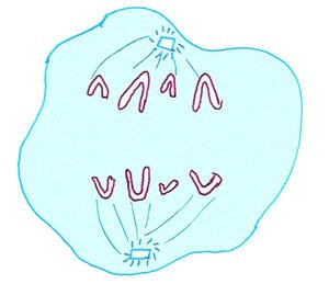
3. Anaphase: Chromatids Separate
Anaphase is the phase where the sister chromatids are separated and pulled to opposite sides of the cell.
- Separation of Sister Chromatids:
The spindle fibers attached to the kinetochores shorten, pulling the sister chromatids apart. Each chromatid is now considered an individual chromosome. - Movement to Opposite Poles:
The chromosomes move toward opposite poles of the cell, guided by the spindle fibers. This movement ensures that each daughter cell will receive an identical set of chromosomes. - Cell Elongation:
Non-kinetochore spindle fibers (those not attached to chromosomes) lengthen, pushing against each other and causing the cell to stretch and elongate.
Anaphase is a critical phase because any errors in chromatid separation can result in aneuploidy-cells with abnormal chromosome numbers-which can cause genetic disorders.
4. Telophase: Nuclear Membranes Reform
Telophase is the phase where the cell starts to reverse the changes seen in prophase, effectively concluding nuclear division.
- Chromosome Decondensation:
The chromosomes, which were tightly coiled for easy movement, begin to decondense back into chromatin, returning to their thread-like structure. - Nuclear Envelope Reformation:
New nuclear membranes form around each set of chromosomes, creating two distinct nuclei within the cell. This reformation ensures that the genetic material is enclosed and protected. - Spindle Fiber Disassembly:
The spindle fibers that were essential for chromosome separation are broken down and recycled for future use.
Telophase marks the end of mitosis, but cell division isn't complete until cytokinesis occurs.
Cytokinesis: Completing Cell Division
Cytokinesis is the process that physically divides the cytoplasm, forming two separate daughter cells. It occurs differently in plant and animal cells:
- In Animal Cells: A cleavage furrow forms, pinching the cell membrane inward until the cell splits.
- In Plant Cells: A cell plate forms at the center, eventually developing into a new cell wall that separates the two daughter cells.
With cytokinesis complete, mitosis ends, and both daughter cells enter interphase, ready for growth and further divisions when needed.
Take This Quiz :

Functions of Mitosis
Mitosis supports various cellular processes beyond growth and repair, ensuring stability, adaptation, and immune defense in living organisms.
- Genetic Stability and Cellular Identity
- Mitosis ensures each daughter cell receives an exact copy of the parent cell's DNA.
- This maintains genetic stability across cell generations, preserving the identity and function of tissues.
- Role in Developmental Patterning (Embryonic Morphogenesis)
- During embryonic development, mitosis helps establish the body plan by regulating cell proliferation in specific regions.
- Controlled mitotic activity shapes tissues, organs, and body structures through processes like pattern formation.
- Clonal Expansion in Adaptive Immunity
- Mitosis supports the adaptive immune response by rapidly producing immune cells when pathogens are detected.
- Activated B-cells and T-cells undergo mitosis to generate clones that target specific infections.
- Wound Healing and Cell Migration
- Mitosis not only replaces damaged cells but also aids in guiding cells to the site of injury.
- During wound healing, cells proliferate and migrate to close the wound, a process involving synchronized mitotic activity.
- Tissue Homeostasis and Turnover
- Mitosis helps maintain tissue homeostasis by balancing cell death (apoptosis) with new cell production.
- This is crucial in tissues with high cell turnover, like the skin, intestinal lining, and blood.
- Mitotic Response to External Stimuli
- Certain external signals, such as growth factors, hormones, and mechanical stress, can trigger mitosis.
- This allows tissues to adapt to changing conditions, such as muscle growth from physical exercise.
Take This Quiz :
Rate this lesson:
 Back to top