Skeletal System: Structure, Functions, and Vital Importance
Lesson Overview
The skeletal system is essential for supporting, protecting, and enabling movement in the body. This lesson will help you understand its structure, functions, and overall importance. Understanding the skeletal system is vital for grasping how the body maintains strength, mobility, and health, as well as protecting vital organs.
What Is the Skeletal System?
The skeletal system is the framework of bones and cartilage that supports and protects the body's vital organs. It provides structure, enables movement, stores minerals, and produces blood cells. In humans, the skeletal system consists of over 200 bones and serves as the body's support structure, helping you stand upright and giving you the ability to move.
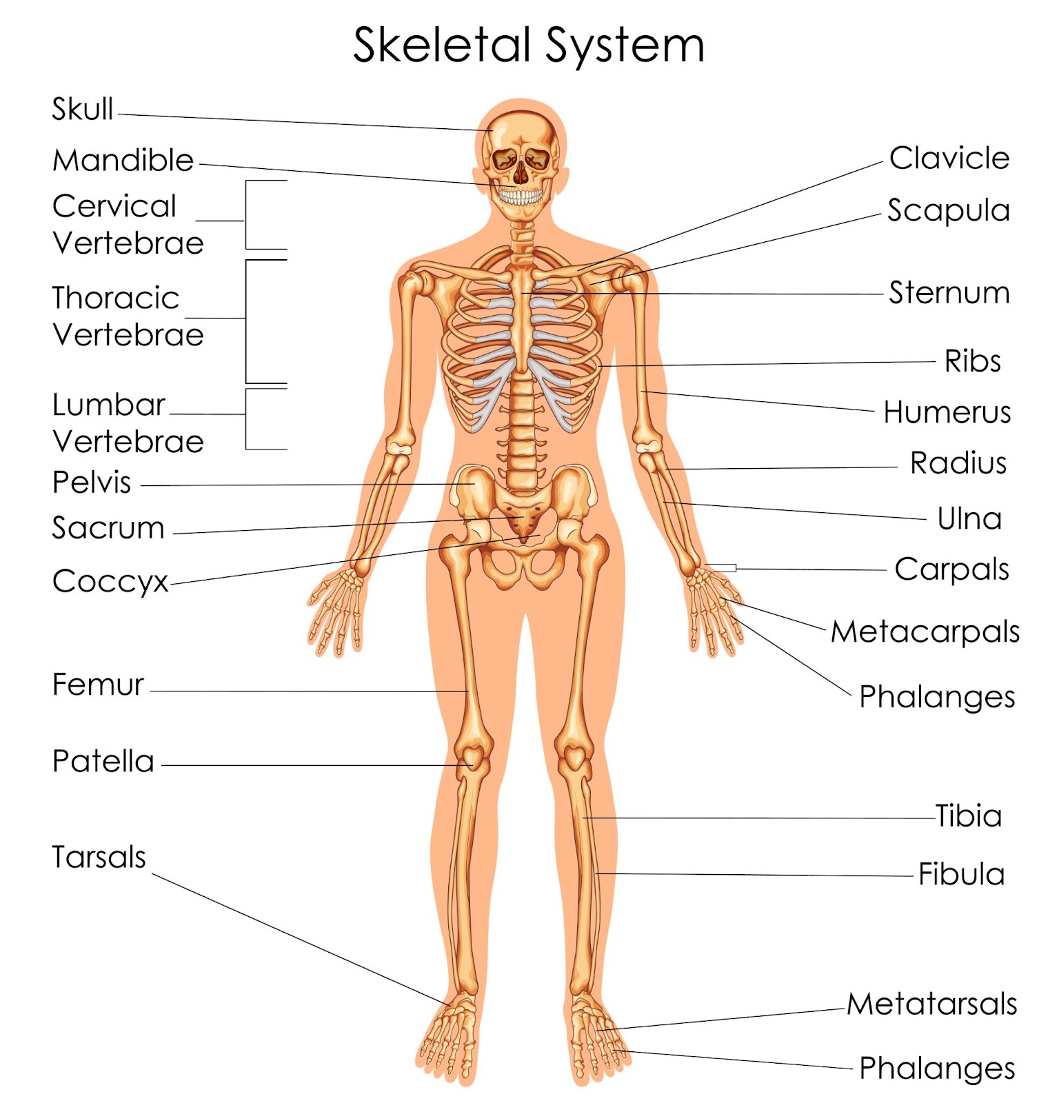
Fig: Human Skeleton Diagram
Human Skeleton Anatomy
The human skeleton consists of a collection of bones and cartilage, which not only provide support but also facilitate movement, protect internal organs, and contribute to essential biological functions like blood production.
The human skeleton anatomy can be broken down into two primary divisions:
1. Appendicular Skeleton
The appendicular skeleton consists of the bones that make up the limbs and the girdles that attach them to the axial skeleton. It includes:
- Upper Limbs: The arms and hands, which are connected to the body via the shoulder girdle. The bones include the humerus, radius, ulna, and various bones in the hands.
- Lower Limbs: The legs and feet, connected to the pelvis. The major bones are the femur, tibia, fibula, and bones of the foot.
- Pelvic Girdle: The hip bones, which support the lower limbs and house the reproductive organs.
- Shoulder Girdle: The collarbones (clavicles) and shoulder blades (scapulae), which help attach the arms to the torso.
2. Axial Skeleton
The axial skeleton forms the central axis of the body, including the skull, vertebral column (spine), and rib cage. It is crucial for protecting the brain, spinal cord, and other vital organs.
- Skull: The skull houses and protects the brain and sensory organs, including the eyes, ears, and nose. It also supports the structures of the face.
- Vertebral Column: The spine consists of vertebrae stacked on top of each other and serves to protect the spinal cord, while providing flexibility and strength for movement.
- Rib Cage: The ribs are connected to the vertebral column and form a protective enclosure around the heart and lungs. They also assist with the breathing process.
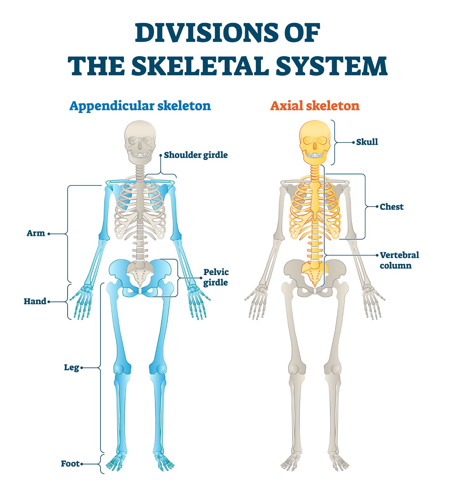
Fig: Divisions of Appendicular and Axial Skeletal System Labeled
Take This Quiz :

How Does the Skeletal System Work?
The skeletal system functions as a complex and coordinated system that supports the body, facilitates movement, protects vital organs, stores minerals, and produces blood cells. By understanding its functions and structure, we can appreciate how the skeletal system function contributes to our overall health.
1. Providing Structure and Support
The skeletal system acts as the body's framework, giving shape and form. Without the skeleton, the body would lack stability and would collapse. The bones of the skeletal system provide a rigid support structure for the muscles, organs, and tissues, helping the body maintain an upright posture.
How It Works:
- The human skeleton anatomy is designed to hold the body upright, distributing weight efficiently so the body can stand and move.
- The human skeleton organs-bones-serve as a solid support for soft tissues like muscles, skin, and internal organs.
2. Facilitating Movement
The skeletal system works in close collaboration with the muscular system to enable movement. Bones act as levers, and muscles act as the motors that make the body move. When muscles contract, they pull on bones, causing movement at the joints.
How It Works:
- Muscles are connected to bones by tendons, and joints are where two bones meet.
- Joints allow bones to move in different ways (e.g., flexion, extension, rotation).
- Ligaments stabilize the joints, ensuring they move in a controlled manner and preventing injury.
3. Protecting Vital Organs
Bones in the skeletal system serve as protective barriers for the body's delicate internal organs. For example, the skull protects the brain, while the ribcage shields the heart and lungs. The spine protects the spinal cord, which is essential for nerve communication throughout the body.
How It Works:
- The bones form a hard casing around the vital organs, keeping them safe from injury.
- The ribcage, composed of ribs and the sternum (breastbone), forms a protective shield for the heart and lungs.
- The skull is the bone structure that houses and protects the brain, one of the most sensitive organs in the body.
4. Storing Minerals
The skeletal system is not only structural but also plays a crucial role in mineral storage. The bones store essential minerals, such as calcium and phosphorus, which are necessary for various bodily functions, including muscle function and nerve signaling.
How It Works:
- When the body requires minerals, they are released from the bones into the bloodstream.
- The human skeleton anatomy acts as a reservoir, ensuring minerals are available when needed to maintain functions like muscle contractions and heart rhythms.
5. Blood Cell Production
The skeletal system produces blood cells through a process known as hematopoiesis. This process primarily occurs in the bone marrow, which is found in the center of certain bones, such as the long bones of the limbs, vertebrae, and pelvis.
How It Works:
- Bone marrow contains stem cells that produce red blood cells (which carry oxygen), white blood cells (which fight infection), and platelets (which help with clotting).
- This production is vital for maintaining a healthy circulatory and immune system.
6. Bone Remodeling and Repair
The skeletal system is dynamic and constantly being remodeled. Bones are broken down and rebuilt throughout life. This process is essential for maintaining bone strength and repairing damage.
How It Works:
- Osteoclasts break down old bone tissue, while osteoblasts create new bone tissue, allowing bones to adapt to stress and injury.
- When a bone fractures, the body initiates the healing process, producing new bone tissue to repair the break.
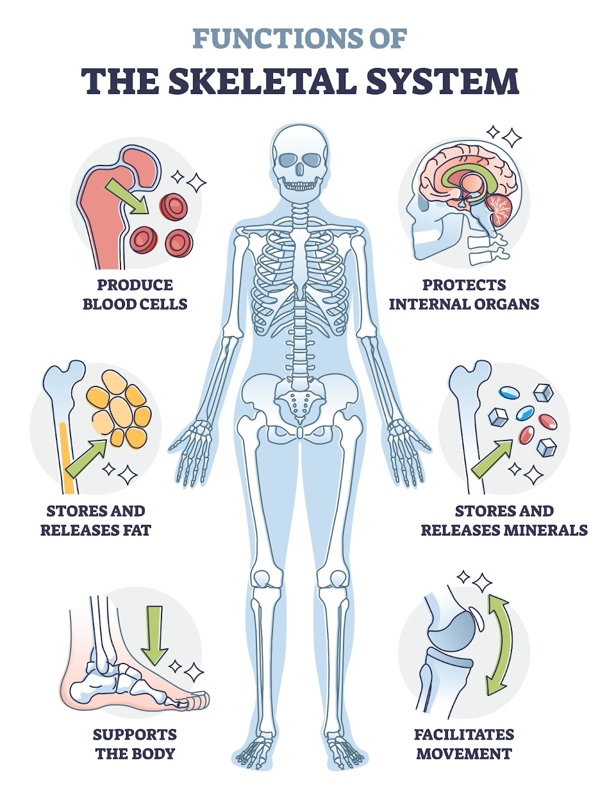
Fig: Skeletal System Functions or Bone Anatomical Functionality Outline Diagram
Components of the Skeletal System
The skeletal system is made up of bones, joints, cartilage, and ligaments, all of which work together to provide structure, support, and mobility to the body. Understanding the components of the skeletal system is crucial in comprehending how the human body functions.
1. Bones
Bones are the primary structural components of the skeletal system, providing the framework that supports the body. There are 206 bones in the adult human body, each varying in size, shape, and function. Bones come in different types, such as long, short, flat, irregular, sesamoid, and sutural bones, each suited for specific roles.
Function:
- Support: Bones give the body its shape and allow for the attachment of muscles and tissues.
- Protection: Bones protect vital organs, such as the brain (skull), heart and lungs (rib cage), and spinal cord (vertebrae).
- Movement: Bones, in conjunction with muscles, facilitate movement.
- Blood Cell Production: The bone marrow inside bones is responsible for producing red blood cells, white blood cells, and platelets.
- Mineral Storage: Bones store essential minerals like calcium and phosphorus, which are released into the bloodstream when needed.
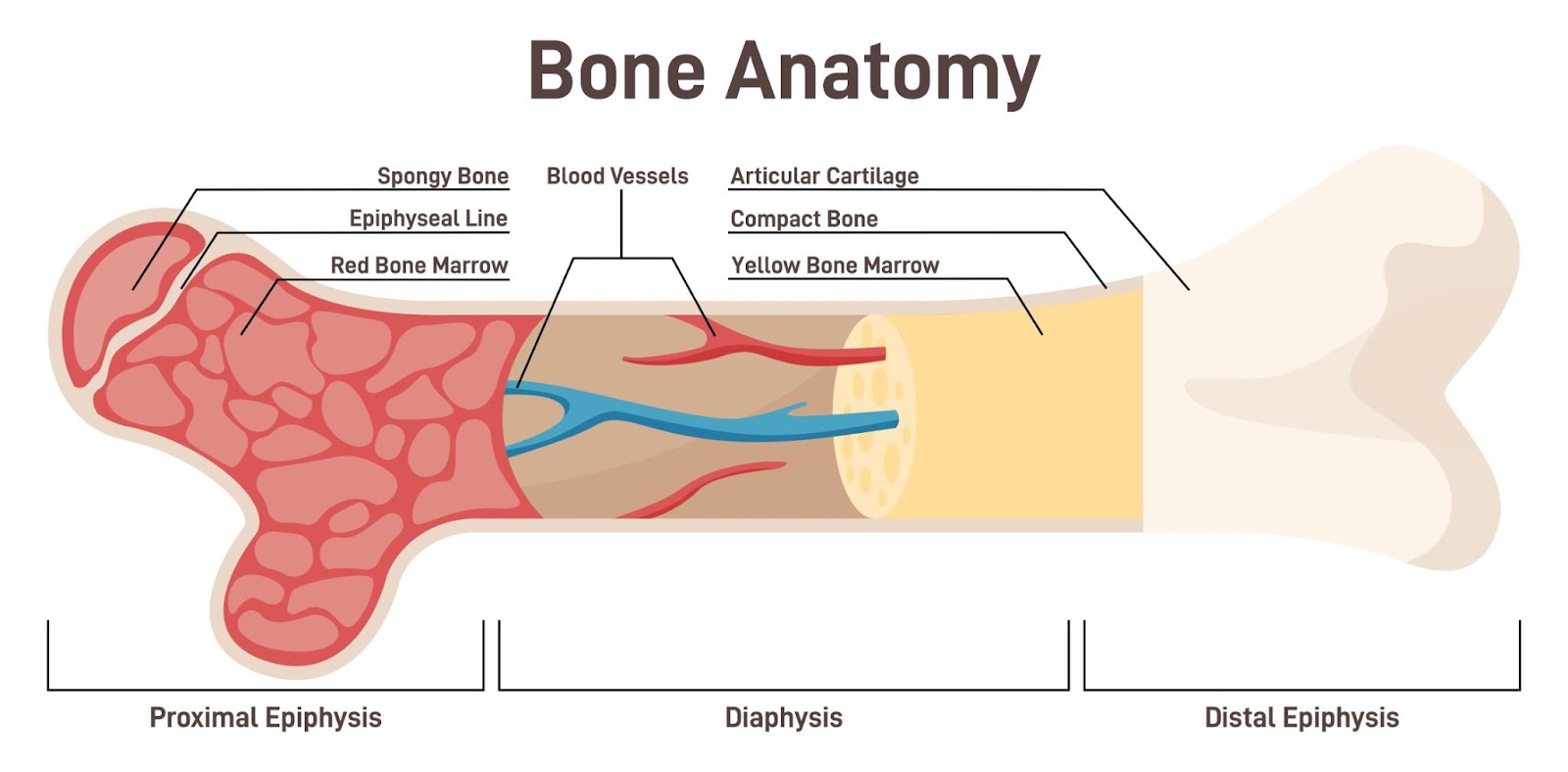
Fig: A detailed anatomical illustration of a bone, labeling the periosteum, endosteum, bone marrow, and trabeculae, showcasing its internal structure and composition.
2. Joints
Joints, also known as articulations, are the areas where two or more bones meet. They allow for movement and flexibility in the body. There are different types of joints based on their structure and the range of movement they allow.
Types of Joints:
- Fibrous Joints: These are immovable joints, such as the sutures in the skull.
- Cartilaginous Joints: These joints allow for limited movement, such as those between the vertebrae of the spine.
- Synovial Joints: These are the most common and allow for a wide range of movements. They include ball-and-socket joints (e.g., shoulder and hip) and hinge joints (e.g., knee and elbow).
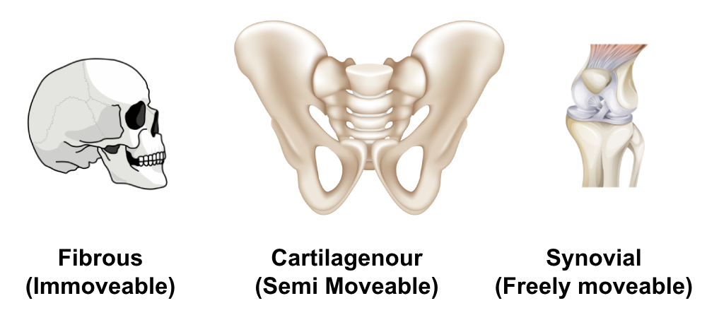
Fig: A diagram showing the types of joints.
Function:
- Movement: Joints enable bones to move in relation to one another.
- Flexibility: They provide flexibility, which allows the body to perform a wide range of motions.
- Shock Absorption: Some joints, such as the knee or ankle, act as shock absorbers to prevent damage during movement.
3. Cartilage
Cartilage is a flexible, smooth tissue that covers the ends of bones at joints. It provides cushioning, reduces friction, and helps with the smooth movement of joints. Cartilage is also found in areas of the body that require flexibility but do not need to be as rigid as bone.
Types of Cartilage:
- Hyaline Cartilage: Found at the ends of long bones, in the ribs, and in the nose.
- Fibrocartilage: Found in intervertebral discs and the pubic symphysis.
- Elastic Cartilage: Found in the ear and the epiglottis.
Function:
- Cushioning and Protection: Cartilage prevents bones from rubbing against each other, reducing friction and wear.
- Flexibility: Cartilage allows for flexible movement in certain parts of the body, like the ribs and ears.
- Shock Absorption: Cartilage helps absorb shock at joints to prevent bone damage.
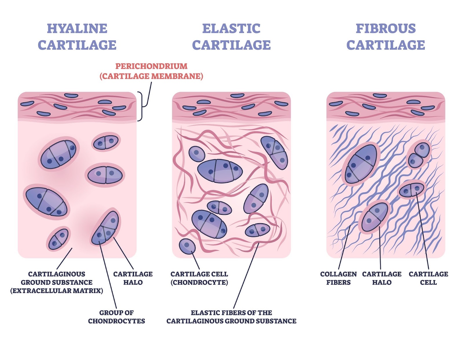
Fig: A labeled diagram showing the types of cartilage-hyaline, fibrous, and elastic-along with the perichondrium membrane.
4. Ligaments
Ligaments are strong, fibrous tissues that connect bones to other bones at joints. They provide stability and limit excessive movement, ensuring the bones stay in proper alignment. Ligaments are an essential part of joint function, as they control the range of motion and help prevent injury.
Function:
- Stabilization: Ligaments stabilize joints, ensuring bones remain in place during movement.
- Prevention of Injury: Ligaments prevent excessive movement, reducing the risk of joint dislocation or strain.
- Support: They provide support to the joints and the entire skeletal structure.
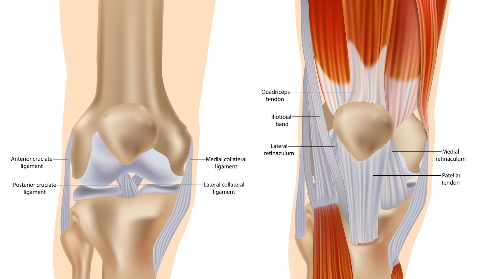
Fig: Knee anatomy including ligaments, cartilage and meniscus.
5. Tendons
Although not technically part of the skeletal system, tendons are closely related to its function. Tendons are strong, fibrous tissues that connect muscles to bones. They play a key role in movement, as they transmit the force generated by muscles to bones.
Function:
- Movement: Tendons enable the movement of bones by transmitting the force exerted by muscles.
- Support: They help muscles remain attached to bones, ensuring proper alignment and function.
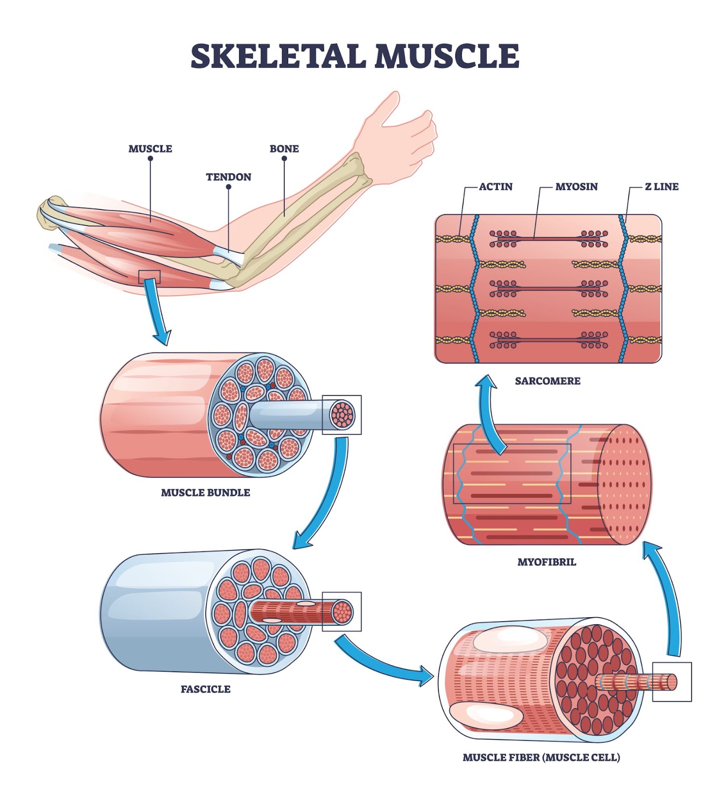
Fig: Skeletal muscle structure layers with anatomical arm closeups outline diagram showing the role of the tendon.
Take This Quiz :

Types of Bones in the Human Body
The skeletal system consists of a variety of bone types, each designed to serve different functions. These bones can be classified based on their shape, structure, and function within the body. Here's a comprehensive look at the different types of bones in the human body and their unique characteristics:
1. Long Bones
Long bones are characterized by their elongated shape, typically longer than they are wide. These bones are primarily found in the limbs and are crucial for movement and supporting body weight.
Examples:
- Femur (thigh bone)
- Humerus (upper arm bone)
- Tibia (shin bone)
- Fibula (calf bone)
Function:
- Long bones are responsible for bearing the weight of the body and providing leverage for movement.
- They contain bone marrow, where blood cells are produced.
2. Short Bones
Short bones are cube-shaped and nearly equal in length, width, and thickness. These bones provide stability and support with little movement, and they often bear weight but are not involved in extensive motion.
Examples:
- Carpals (wrist bones)
- Tarsals (ankle bones)
Function:
- Short bones provide support and help in absorbing shock, as they allow limited movement.
- They are typically found in the wrists, ankles, and hands, offering stability to joints.
3. Flat Bones
Flat bones have a thin, flat shape and provide protection to internal organs. They also offer a large surface area for muscle attachment.
Examples:
- Skull bones (such as the frontal bone and parietal bones)
- Ribs
- Sternum (breastbone)
- Scapulae (shoulder blades)
Function:
- Flat bones are primarily designed for protection, safeguarding the brain, heart, and lungs.
- These bones also serve as a surface for muscle attachment, enabling movement.
4. Irregular Bones
Irregular bones have complex shapes that do not fit into the categories of long, short, or flat bones. These bones serve various purposes in the body, from providing protection to enabling movement.
Examples:
- Vertebrae (bones of the spine)
- Facial bones (such as the mandible or jawbone)
- Pelvis bones
Function:
- Irregular bones protect internal organs, such as the spinal cord and certain parts of the brain.
- They also serve as attachment points for muscles and ligaments, contributing to overall movement.
5. Sesamoid Bones
Sesamoid bones are small, round bones embedded within tendons. These bones help reduce friction and pressure on tendons during movement and provide leverage for muscles.
Examples:
- Patella (kneecap)
- Small sesamoid bones found in the hands and feet
Function:
- Sesamoid bones protect tendons from stress and wear by providing a smooth surface for tendons to slide over.
- They also increase the mechanical advantage of muscles, making movement more efficient.
6. Accessory or Sutural Bones
These are extra bones that can form in the sutures (joints) between flat bones of the skull. They are not present in every individual but can appear as extra ossifications in certain regions of the skull.
Examples:
- Sutural bones in the skull (e.g., between the parietal and occipital bones)
Function:
- Sutural bones are considered non-essential, but they may contribute to the overall structure and strength of the skull.
- Their function is not fully understood but may involve reinforcing skull joints.
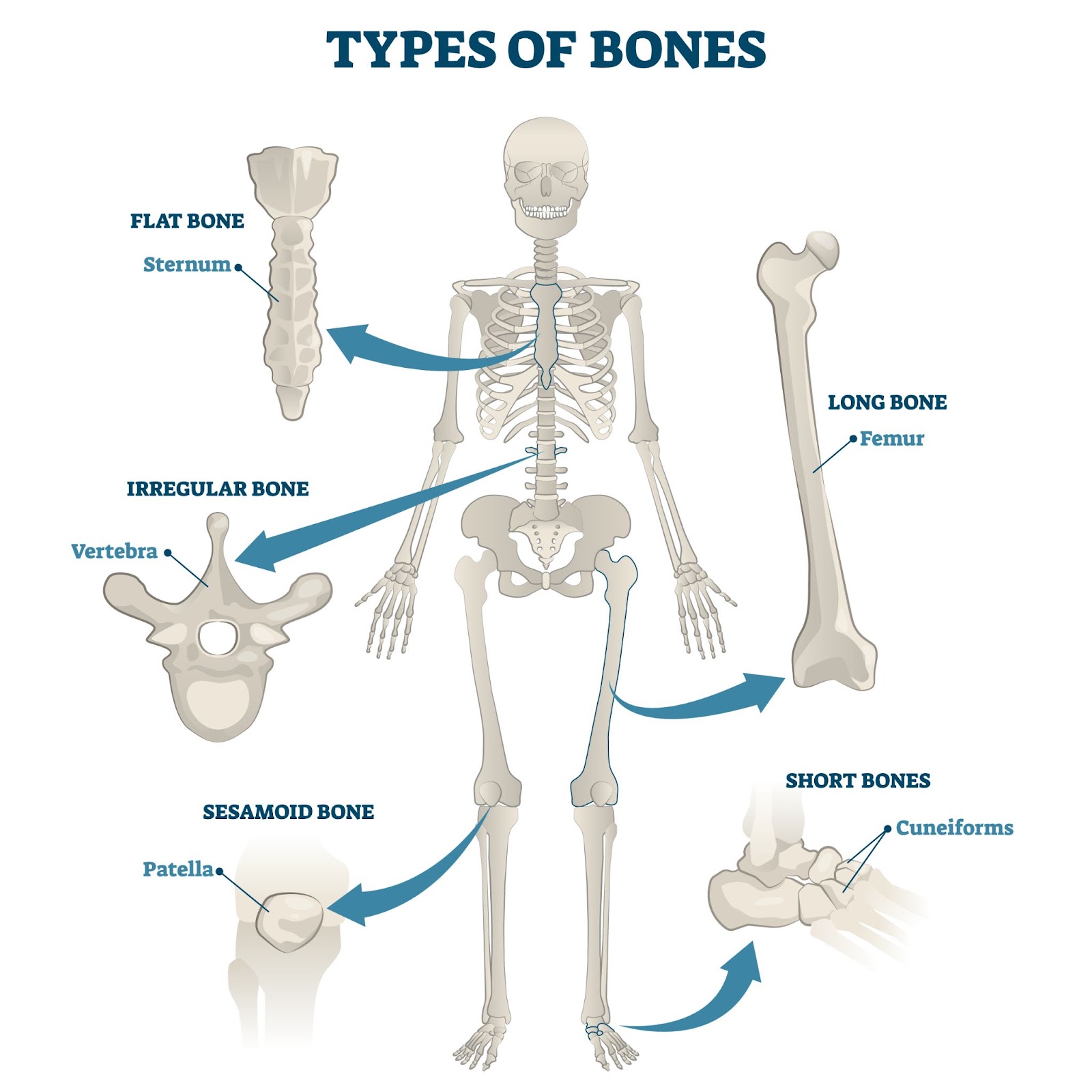
Fig: A Diagram Showing Types of Bones
Rate this lesson:
 Back to top